Biomod/2014/OhioMOD/experimentnotes
<html> <head> <script src="http://ajax.googleapis.com/ajax/libs/jquery/1.11.1/jquery.min.js"></script> <script src="http://maxcdn.bootstrapcdn.com/bootstrap/3.2.0/js/bootstrap.min.js"></script> <link href="http://maxcdn.bootstrapcdn.com/bootswatch/3.2.0/yeti/bootstrap.min.css" rel="stylesheet"> <style>
- content{margin-left:0;}
.navbar{ top:8.5px; position:fixed; width:96.8%; } /*clearing the side openwetware panels*/ .firstHeading, #column-one, #p-bookmarks, #p-history , .portlet{ display:none; }
- bodyContent/*clearing the side openwetware panels
.firstHeading, #column-one, #p-bookmarks, #p-history , .portlet{ display:none; }
- content#bodyContent, {
/*display:none;*/ background-color:#FFFFFF; }
- OSUfooter{
clear:both; }
- footer{display: none;}
/*Done clearing*/
</style>
</head>
</html> <html> <head> <style> center {padding-bottom:50px;}
- ExperimentNotes{
 width: 700px ;
 margin-left: auto ;
 margin-right: auto ;
 margin-bottom: 50px;
}
- list2
{
text-indent:50px;
} </style> <style type="text/css"> p{text-indent:50px;} </style>
</head> <body>
Experiment Notes
| Table of Contents | |
|---|---|
|
1. Cellular Uptake Experiment (DNA Extraction)v1
|
|
1.Cellular Uptake Experiment (DNA Extraction) v1
07/29/2014
Materials:
Procedure:
- The concentration was 1.5 nM.
- Final volume: 250 uL
- Final concentration of structures: 0.3 nM
07/30/2014
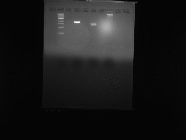
Results: <figure>
<img src="http://openwetware.org/images/e/e5/Gel_verification.jpg"height="300" width="300"/> <figcaption>Fig: Gel verification of DNA extraction. From left to right: ladder, scaffold, purified branch structure, DNA extract.</figcaption> </figure>
As the gel shows, the DNA extract had no band that corresponded with the branch purified structure.
Discussion:
Since there was no band on the gel corresponding with the folded branch structures, there is no way to confirm whether the structures were uptaken or not and whether the DNA extraction was able to extract the DNA origami structures or not. If structures were uptaken, then the concentration of structures might have been too dilute to register on the gel, since the initial concentration in solution was only 0.3 nM.
For future experiments, the cells should be incubated in a higher concentration of structures. In addition, TEM imaging can be used to determine whether there are any structures present in the DNA extracted from the cell.
<a href="http://openwetware.org/index.php?title=Biomod/2014/OhioMOD/experimentnotes&action=edit">Edit Experiment Notes</a>
</body>
</html>
<html>
<head> <style>
- Footer table {
  width: 96.8%;
  max-width:2350px;
  bottom:0px;
  height:50px;   /* Height of the footer */
  font-weight:300;
  background-color: #2E2E2E;
   text-align:center;
   color: white;
   font-size:13px;
   border: 3px white solid;
}
- Footer {
   clear: both; /*may be omitted*/
   position: absolute;
   bottom: 12px;
   background-color:#fffff;
   width: 100%;
   height: 40px; /* or anything you like */
}
</style>
</head>
</html>
