Biomod/2013/Todai/Experiment: Difference between revisions
Kenta Koyama (talk | contribs) No edit summary |
Kenta Koyama (talk | contribs) No edit summary |
||
| Line 192: | Line 192: | ||
<h1 class="title"><a name="PilotStudy"> Pilot Study</a></h1> | <h1 class="title"><a name="PilotStudy"> Pilot Study</a></h1> | ||
<!-- | <!--Assembly of DNA structures--> | ||
<article> | <article> | ||
| Line 209: | Line 209: | ||
<figure> | <figure> | ||
<center> | <center> | ||
<img src="http://openwetware.org/images/ | <img src="http://openwetware.org/images/7/71/640px_suv_dls_popg50r60-Todai.png" width=640px height=360px > | ||
<figcaption> <b>The result of DLS </b> | <figcaption> <b>The result of DLS (Viscotek, 802 DLS)</b> | ||
</figcaption> | </figcaption> | ||
</center> | </center> | ||
</figure> | </figure> | ||
<div class="zairyou-heading">[Discussion]</div> | <div class="zairyou-heading">[Discussion]</div> | ||
<p class="paragraph"> | <p class="paragraph">For floating assay, uniformly-sized liposome were prepared. DLS data shows sharp peak with the mean radius of 60 nm, indicating the homogenity of liposomes. | ||
</p> | </p> | ||
</article> | </article> | ||
| Line 227: | Line 224: | ||
<article> | <article> | ||
<div class="mini-title"> | <div class="mini-title"> | ||
<a name="Flotation_assay_of_liposome_and_DNA_origami">2-2. Floatation assay of liposome and DNA origami</a> | <a name="Flotation_assay_of_liposome_and_DNA_origami">2-2. Floatation assay of liposome and Rectangular tile(DNA origami)</a> | ||
<figure> | <figure> | ||
<center> | <center> | ||
| Line 253: | Line 250: | ||
retrieved from supermetant liquid and a sample(fraction 6) from precipitation by the addition of buffer used in | retrieved from supermetant liquid and a sample(fraction 6) from precipitation by the addition of buffer used in | ||
assay. When the sample, tile mixed with liposomes, were assayed, tiles were observed in the top layer. The distribut ion of liposomes is observed by the fluorescence of NIL(Nile Red). | assay. When the sample, tile mixed with liposomes, were assayed, tiles were observed in the top layer. The distribut ion of liposomes is observed by the fluorescence of NIL(Nile Red). | ||
</p> | |||
<br> | |||
</article> | |||
<!--hybridization of Cholesterol Oligo with OCK--> | |||
<article> | |||
<div class="mini-title"> | |||
<a name="hybridization_of_Cholesterol_Oligo_with_OCK">hybridization of Cholesterol Oligo with OCK</a> | |||
<figure> | |||
<center> | |||
<img src="http://openwetware.org/images/d/d7/480px_OCKchol-Todai.png" width=480px height=300px > | |||
</center> | |||
</figure> | |||
<br> | |||
<div class="zairyou-heading">[Discussion]</div> | |||
<p class="paragraph"> | |||
The result of 1 % agarose gel electrophoresis showed that the band of | |||
sample 5 and 7 were smeared, showing the successful of hybridization of | |||
cholesterol oligo with OCK (Langecker et al. (2012)). We concluded that | |||
the optimized condition for hybridization is: 1 hour incubation at room | |||
temperature with 5 times excess cholesterol oligo to OCK. | |||
</p> | </p> | ||
<br> | <br> | ||
| Line 420: | Line 442: | ||
</article> | </article> | ||
<br> | <br> | ||
<!--Research for azobenzene--> | |||
<article> | |||
<div class="mini-title"> | |||
<a name="Transmission_electron_microscopy"> | |||
Transmission electron microscopy(TEM) | |||
</a> | |||
</div> | |||
<!--Procedure--> | |||
<div class="zairyou-heading">[Procedure]<sup>[6]</sup></div> | |||
<p class="paragraph"> | |||
The procedure of TEM was refered to previous researches<sup>[6]</sup>. | |||
</p> | |||
</article> | |||
<!--liposome--> | <!--liposome--> | ||
<article> | <article> | ||
<h2>2.Flotation assay</h2> | <h2>2.Flotation assay</h2> | ||
<div class="mini-title"> | |||
<a name=" | <a name="Preparation_of_SUVs">Preparation of SUVs</a> | ||
</div> | |||
<h3>Type 1: POPC 100%</h3> | |||
<!--Reagent--> | <!--Reagent--> | ||
<div class="zairyou-heading">[Reagent]</div> | <div class="zairyou-heading">[Reagent]</div> | ||
| Line 472: | Line 510: | ||
</article> | </article> | ||
<br> | <br> | ||
<!--Preparation of SUVs_Added--> | |||
<article> | |||
<h3>Type 2: POPC 50%, POPG 50%</h3> | |||
<!--Reagent--> | |||
<div class="zairyou-heading">[Reagent]</div> | |||
<br> | |||
<table> | |||
<tr> | |||
<th>Lipid mix<sup>*</sup></th> | |||
<td>3 ml</td> | |||
</tr> | |||
<tr> | |||
<th>150 mM aqueous KCl solution</th> | |||
<td>1 µL</td> | |||
</tr> | |||
</table> | |||
<br> | |||
*...Lipid mix | |||
<table> | |||
<tr> | |||
<th>5 mg/mL POPC</th> | |||
<td>0.1 mL</td> | |||
</tr> | |||
<tr> | |||
<th>5 mg/mL POPG</th> | |||
<td>0.1 mL</td> | |||
</tr> | |||
<tr> | |||
<th>10 uM NileRed solution<sub>2</sub></th> | |||
<td>0.13 mL</td> | |||
</tr> | |||
<tr> | |||
<th>Chloroform</th> | |||
<td>2.67 mL</td> | |||
</tr> | |||
</table> | |||
<!-- | <!--Procedure--> | ||
<div class="zairyou-heading">[Procedure]</div> | |||
<ul class="procedure-list"> | |||
<li>A lipid film was formed by evaporating 3 ml of lipid mix in a 50 ml | |||
eggplant flask, using a rotational evaporator (EYELA, model#N1110) for | |||
10 mins. | |||
</li> | |||
<li>The flask was kept under vacuum overnight to evaporate remaining chloroform.</li> | |||
<li>The lipid film was resuspended in 1 mL of 150 mM aqueous KCl solution.</li> | |||
<li>Lipid suspended solution was filtered through 100nm polar filter using extruder (Avanti) to prepare uniformly-sized liposome.</li> | |||
<li>The size of liposome was measured with DLS (Viscotek 802 DLS).</li> | |||
<li>The solution was kept at 3℃ until usage.</li> | |||
</ul> | |||
</article> | |||
<br> | |||
<!--Preparation_of_GUVs_Added--> | |||
<article> | <article> | ||
<div class="mini-title"> | <div class="mini-title"> | ||
<a name=" | <a name="Preparation_of_GUVs">Preparation of GUVs</a> | ||
</div> | </div> | ||
<!--Reagent--> | |||
<div class="zairyou-heading">[Reagent]</div> | |||
<br> | |||
<table> | |||
<tr> | |||
<th>Lipid mix<sup>*</sup></th> | |||
<td>3 ml</td> | |||
</tr> | |||
<tr> | |||
<th>150 mM aqueous KCl solution</th> | |||
<td>1 µL</td> | |||
</tr> | |||
</table> | |||
<br> | |||
*...Lipid mix | |||
<table> | |||
<tr> | |||
<th>5 mg/mL POPC</th> | |||
<td>0.1 mL</td> | |||
</tr> | |||
<tr> | |||
<th>5 mg/mL POPG</th> | |||
<td>0.1 mL</td> | |||
</tr> | |||
<tr> | |||
<th>10 uM NileRed solution<sub>2</sub></th> | |||
<td>0.13 mL</td> | |||
</tr> | |||
<tr> | |||
<th>Chloroform</th> | |||
<td>2.67 mL</td> | |||
</tr> | |||
</table> | |||
<!--Procedure--> | |||
<div class="zairyou-heading">[Procedure]</div> | |||
<ul class="procedure-list"> | |||
<li>A lipid film was formed by evaporating 3 ml of lipid mix in a 50 ml | |||
eggplant flask, using a rotational evaporator (EYELA, model#N1110) for | |||
10 mins. | |||
</li> | |||
<li>The flask was kept under vacuum overnight to evaporate remaining chloroform.</li> | |||
<li>The lipid film was resuspended in 1 mL of 150 mM aqueous KCl solution.</li> | |||
</ul> | |||
</article> | |||
<br> | |||
<!--flotation assay_editted--> | |||
<!--Protocol Hybridization of cholesterol oligo with OCK_Added--> | |||
<article> | |||
<div class="mini-title"> | |||
<a name="Protocol_Hybridization_of_cholesterol_oligo_with_OCK">Hybridization of cholesterol oligo with OCK</a> | |||
</div> | |||
<!--Reagent--> | <!--Reagent--> | ||
<div class="zairyou-heading">[Reagent]</div> | <div class="zairyou-heading">[Reagent]</div> | ||
<br> | |||
<table> | <table> | ||
<tr> | <tr> | ||
<th> | <th>OCK</th> | ||
<td> | <td>48 µL</td> | ||
</tr> | </tr> | ||
<tr> | <tr> | ||
<th> | <th>Cholesterol oligo (0.32, 0.64, 3.2, 6.4 µM)</th> | ||
<td> | <td>100 µL</td> | ||
</tr> | </tr> | ||
</table> | |||
<center> | |||
Marker; GeneRuler DNA Ladder Mix (Fermentas, GeneRuler DNA Ladder Mix #SM0331) | |||
</center> | |||
<!--Procedure--> | |||
<div class="zairyou-heading">[Procedure]</div> | |||
<ul class="procedure-list"> | |||
<li>Centrifuge for 16 minutes at 100 krpm at 4 ℃ using TLA 100.2 rotor (BECKMAN COULTER) with Ultracentrifuge (BECKMAN COULTER, Optima MAX-XP).</li> | |||
<li>Each sample was mixed and incubated as shown below:<sup>*</sup></li> | |||
</table> | |||
<br> | |||
*...Table1. | |||
<table> | |||
<center> | |||
<tr> | |||
<th>Sample No.</th> | |||
<td>1</td> | |||
<td>2</td> | |||
<td>3</td> | |||
<td>4</td> | |||
<td>5</td> | |||
<td>6</td> | |||
<td>7</td> | |||
<td>8</td> | |||
</tr> | |||
<tr> | <tr> | ||
<th> | <th>Purified OCK (40 µM)</th> | ||
<td> | <td>6 µL</td> | ||
<td>6 µL</td> | |||
<td>6 µL</td> | |||
<td>6 µL</td> | |||
<td>6 µL</td> | |||
<td>6 µL</td> | |||
<td>6 µL</td> | |||
<td>6 µL</td> | |||
</tr> | |||
<tr> | |||
<th>0.32 µM Cholesterol oligo</th> | |||
<td>1.5 µL</td> | |||
<td>1.5 µL</td> | |||
<td>-</td> | |||
<td>-</td> | |||
<td>-</td> | |||
<td>-</td> | |||
<td>-</td> | |||
<td>-</td> | |||
</tr> | |||
<tr> | |||
<th>0.64 µM Cholesterol oligo</th> | |||
<td>-</td> | |||
<td>-</td> | |||
<td>1.5 µL</td> | |||
<td>1.5 µL</td> | |||
<td>-</td> | |||
<td>-</td> | |||
<td>-</td> | |||
<td>-</td> | |||
</tr> | </tr> | ||
<tr> | <tr> | ||
<th> | <th>3.2 µM Cholesterol oligo</th> | ||
<td> | <td>-</td> | ||
<td>-</td> | |||
<td>-</td> | |||
<td>-</td> | |||
<td>1.5 µL</td> | |||
<td>1.5 µL</td> | |||
<td>-</td> | |||
<td>-</td> | |||
</tr> | |||
<tr> | |||
<th>6.4 µM Cholesterol oligo</th> | |||
<td>-</td> | |||
<td>-</td> | |||
<td>-</td> | |||
<td>-</td> | |||
<td>-</td> | |||
<td>-</td> | |||
<td>1.5 µL</td> | |||
<td>1.5 µL</td> | |||
</tr> | |||
<tr> | |||
<th>[Cholesterol oligo]/[OCK] (see #Note)</th> | |||
<td>1/2</td> | |||
<td>1/2</td> | |||
<td>1</td> | |||
<td>1</td> | |||
<td>5</td> | |||
<td>5</td> | |||
<td>10</td> | |||
<td>10</td> | |||
</tr> | |||
<tr> | |||
<th>Incubation time [min]</th> | |||
<td>60</td> | |||
<td>30</td> | |||
<td>60</td> | |||
<td>30</td> | |||
<td>60</td> | |||
<td>30</td> | |||
<td>60</td> | |||
<td>30</td> | |||
</tr> | </tr> | ||
</center> | |||
</table> | </table> | ||
#Note; OCK has 4 cholesterol oligo binding sites. Therefore, we devided the molar ratio of cholesterol oligo to OCK with 4. | |||
<li>Each sample was analyzed by 1% agarose gel electrophoresis (100V, 1 hour).</li> | |||
</ul> | |||
</article> | |||
<br> | |||
<article> | |||
<!--Flotation assay(OCK)_Added--> | |||
<article> | |||
<div class="mini-title"> | |||
<a name="Flotation_assay_[OCK]">Flotation assay [OCK]</a> | |||
</div> | |||
<!--Reagent--> | |||
<div class="zairyou-heading">[Reagent]</div> | |||
<br> | |||
<table> | <table> | ||
<tr> | <tr> | ||
<th> | <th>OCK</th> | ||
<td> | <td>100 µL</td> | ||
</tr> | |||
<tr> | |||
<th>Cholesterol hybridized OCK</th> | |||
<td>100 µL</td> | |||
</tr> | |||
<tr> | |||
<th>Liposome (1 mg/mL SUVs)</th> | |||
<td>100 µL</td> | |||
</tr> | </tr> | ||
<tr> | <tr> | ||
<th> | <th>2.25 M Sucrose buffer<sup>*</sup></th> | ||
<td> | <td>500 µL</td> | ||
</tr> | </tr> | ||
<tr> | <tr> | ||
<th> | <th>1.6 M Sucrose buffer<sup>**</sup></th> | ||
<td> | <td>900 µL</td> | ||
</tr> | </tr> | ||
<tr> | <tr> | ||
<th> | <th>150 mM aqueous KCl solution</th> | ||
<td> | <td>100 µL</td> | ||
</tr> | </tr> | ||
<tr> | <tr> | ||
<th> | <th>1×Flotation buffer<sup>**</sup></th> | ||
<td> | <td>600 µL</td> | ||
</tr> | </tr> | ||
</table> | |||
<br> | |||
*...2.25 M Sucrose buffer | |||
<table> | |||
<tr> | |||
<th>HEPES-KOH (pH 7.6)</th> | |||
<td>50 mM</td> | |||
</tr> | |||
<tr> | <tr> | ||
<th>KCl</th> | <th>KCl</th> | ||
<td> | <td>100 mM</td> | ||
</tr> | |||
<tr> | |||
<th>MgCl<sub>2</sub></th> | |||
<td>20 mM</td> | |||
</tr> | </tr> | ||
<tr> | |||
<th>Sucrose</th> | |||
<td>2.25 M</td> | |||
</tr> | |||
</table> | |||
</table> | |||
<br> | |||
**...1.6 M Sucrose buffer | |||
<table> | |||
<tr> | <tr> | ||
<th>HEPES-KOH (pH 7.6)</th> | |||
<td>50 mM</td> | |||
</tr> | |||
<tr> | |||
<th>KCl</th> | |||
<td>100 mM</td> | |||
</tr> | |||
<tr> | |||
<th>MgCl<sub>2</sub></th> | <th>MgCl<sub>2</sub></th> | ||
<td> | <td>20 mM</td> | ||
</tr> | </tr> | ||
<tr> | <tr> | ||
<th>Sucrose</th> | <th>Sucrose</th> | ||
<td>1. | <td>1.6 M</td> | ||
</tr> | </tr> | ||
</table> | </table> | ||
</table> | |||
<br> | <br> | ||
***...1×Flotation buffer | |||
<table> | |||
<tr> | |||
<th>HEPES-KOH (pH 7.6)</th> | |||
<td>50 mM</td> | |||
</tr> | |||
<tr> | |||
<th>KCl</th> | |||
<td>100 mM</td> | |||
</tr> | |||
<tr> | |||
<th>MgCl<sub>2</sub></th> | |||
<td>20 mM</td> | |||
</tr> | |||
</table> | |||
<!--Procedure--> | <!--Procedure--> | ||
<div class="zairyou-heading">[Procedure]</div> | <div class="zairyou-heading">[Procedure]</div> | ||
<ul class="procedure-list"> | <ul class="procedure-list"> | ||
<li>sample | <li>Each sample was mixed as shown below:<sup>****</sup></li> | ||
<li> | </table> | ||
<li> | <br> | ||
<li> | ****...Table1. Breakdown of Samples | ||
<li> | <table> | ||
<tr> | |||
<th>Sample No.</th> | |||
<td>1</td> | |||
<td>2</td> | |||
<td>3</td> | |||
<td>4</td> | |||
</tr> | |||
<tr> | |||
<th>OCK</th> | |||
<td>50 µL</td> | |||
<td>50 µL</td> | |||
<td>-</td> | |||
<td>-</td> | |||
</tr> | |||
<tr> | |||
<th>Cholesterol hybridized OCK</th> | |||
<td>-</td> | |||
<td>-</td> | |||
<td>50 µL</td> | |||
<td>50 µL</td> | |||
</tr> | |||
<tr> | |||
<th>Liposome</th> | |||
<td>50 µL</td> | |||
<td>-</td> | |||
<td>50 µL</td> | |||
<td>-</td> | |||
</tr> | |||
<tr> | |||
<th>150 mM aqueous KCl solution</th> | |||
<td>-</td> | |||
<td>50 µL</td> | |||
<td>-</td> | |||
<td>50 µL</td> | |||
</tr> | |||
<tr> | |||
<th>2.25 M Sucrose buffer</th> | |||
<td>125 µL</td> | |||
<td>125 µL</td> | |||
<td>125 µL</td> | |||
<td>125 µL</td> | |||
</tr> | |||
</table> | |||
<li>225 µL of 1.6 M sucrose buffer was overlaid with 225 µL of sample | |||
mixture in centrifuge tubes (Beckman, cat#343778, 11 x 34 mm). | |||
</li> | |||
<li>Centrifuge for 16 minutes at 100 krpm at 4 ℃ using TLA 100.2 rotor (BECKMAN COULTER) with Ultracentrifuge (BECKMAN COULTER, Optima MAX-XP).</li> | |||
<li>150 µL of supernatant was extracted from top to bottom for 3 times (Fraction 1 to 3) and the pellet was retrieved with 150 µL of 1×Flotation buffer (Fraction 4).</li> | |||
<li>Fraction 1-4 of each sample were analyzed by 1 % agaraose gel | |||
electrophoresis (100V, 1 hour). | |||
</li> | |||
<li>The Intensity of fluorescence of NileRed (Liposome) was measured with fluorescence spectrophotometer (JASCO, FP-6500) to investigate the existence of liposome in each Fraction.</li> | |||
<li>The radiuses of liposome of each fraction were measured with DLS (Viscotek, 802 DLS).</li> | |||
</ul> | </ul> | ||
</article> | </article> | ||
<br> | <br> | ||
<br> | |||
</article> | |||
<article> | <article> | ||
| Line 686: | Line 1,054: | ||
</article> | </article> | ||
<br> | <br> | ||
<!--Accelerated Click reaction_Added--> | |||
<article> | |||
<div class="mini-title"> | |||
<a name="Accelerated_Click_reaction">Accelerated Click reaction (using streptavidin to make the aklyne and azide reactive groups close) </a> | |||
</div> | |||
<!--Reagent--> | |||
<div class="zairyou-heading">[Reagent]</div> | |||
<br> | |||
<table> | |||
<tr> | |||
<th>2x barrel buffer</th> | |||
<td>6 µL</td> | |||
</tr> | |||
<tr> | |||
<th>alkyne oligo (carrying biotin) (15 µM)</th> | |||
<td>1 µL</td> | |||
</tr> | |||
<tr> | |||
<th>azide oligo (carrying biotin) (15 µM)</th> | |||
<td>1 µL</td> | |||
</tr> | |||
<tr> | |||
<th>streptavidin (500 µM)</th> | |||
<td>1 µL</td> | |||
</tr> | |||
</table> | |||
<br> | |||
<!--Procedure--> | |||
<div class="zairyou-heading">[Procedure]</div> | |||
<ul class="procedure-list"> | |||
<li>mix reagents</li> | |||
<li>incubate the tube at 37 ℃ for indicated reaction time.</li> | |||
<li>boil at 95 ℃ for 30 minutes to break down streptavidin</li> | |||
</ul> | |||
</article> | |||
<br> | |||
<!--Click reaction (using hybridization to make the aklyne and azide reactive groups close)_Added--> | |||
<article> | |||
<div class="mini-title"> | |||
<a name="Click_reaction_(using_hybridization_to_make_the_aklyne_and_azide_reactive_groups close)">Click reaction (using hybridization to make the aklyne and azide reactive groups close) </a> | |||
</div> | |||
<!--Reagent--> | |||
<div class="zairyou-heading">[Reagent]</div> | |||
<br> | |||
<table> | |||
<tr> | |||
<th>2x barrel buffer</th> | |||
<td>7 µL</td> | |||
</tr> | |||
<tr> | |||
<th>alkyne oligo (15 µM)</th> | |||
<td>1 µL</td> | |||
</tr> | |||
<tr> | |||
<th>azide oligo (15 µM)</th> | |||
<td>1 µL</td> | |||
</tr> | |||
<tr> | |||
<th>scaffold (15 µM)</th> | |||
<td>1 µL</td> | |||
</tr> | |||
</table> | |||
<br> | |||
<!--Procedure--> | |||
<div class="zairyou-heading">[Procedure]</div> | |||
<ul class="procedure-list"> | |||
<li>mix reagents</li> | |||
<li>incubate the tube at 37 ℃.</li> | |||
<li>add loading buffer into the reaction mixture and boil at 95 ℃ for 5 minutes to denature the double strand to single strand.</li> | |||
</ul> | |||
</article> | |||
<br> | |||
<!--Click reaction (copper catalyst-free)_Added--> | |||
<div class="mini-title"> | |||
<a name="Click_reaction_(copper_catalyst-free)">Click reaction (copper catalyst-free)</a> | |||
</div> | |||
<!--Reagent--> | |||
<div class="zairyou-heading">[Reagent]</div> | |||
<br> | |||
<table> | |||
<tr> | |||
<th>2x barrel buffer</th> | |||
<td>7 µL</td> | |||
</tr> | |||
<tr> | |||
<th>alkyne oligo (15 µM)</th> | |||
<td>1 µL</td> | |||
</tr> | |||
<tr> | |||
<th>azide oligo (15 µM)</th> | |||
<td>1 µL</td> | |||
</tr> | |||
<tr> | |||
<th>scaffold (15 µM)</th> | |||
<td>1 µL</td> | |||
</tr> | |||
</table> | |||
<br> | |||
<!--※10x OCK buffer (f. 100 µl)--> | |||
<div class="zairyou-heading">[※※ 2x barrel buffer]</div> | |||
<br> | |||
<table> | |||
<tr> | |||
<th>1M Tris (pH 7.5)</th> | |||
<td>5 µL</td> | |||
</tr> | |||
<tr> | |||
<th>0.5M EDTA</th> | |||
<td>2 µL</td> | |||
</tr> | |||
<tr> | |||
<th>5M NaCl</th> | |||
<td>1 µL</td> | |||
</tr> | |||
<tr> | |||
<th>MQ</th> | |||
<td>32 µL</td> | |||
</tr> | |||
</table> | |||
<br> | |||
<!--Procedure--> | |||
<div class="zairyou-heading">[Procedure]</div> | |||
<ul class="procedure-list"> | |||
<li>mix reagents</li> | |||
<li>incubate the tube at 37 ℃.</li> | |||
<li>add loading buffer into the reaction mixture and boil at 95 ℃ for 5 minutes to denature the double strand to single strand.</li> | |||
</ul> | |||
</article> | |||
<br> | |||
<!--Research for azobenzene--> | <!--Research for azobenzene--> | ||
| Line 776: | Line 1,316: | ||
<div class="reference-journal"> | <div class="reference-journal"> | ||
Angewandte Chemie International Edition,2009,48(37),6820–6823 | Angewandte Chemie International Edition,2009,48(37),6820–6823 | ||
</div> | |||
</div> | |||
<div> | |||
<div class="reference-title"> | |||
<a name="proref-1"> | |||
[6] A primer to scaffolded DNA origami.</a> | |||
</div> | |||
<div class="reference-author"> | |||
Castro CE, Kilchherr F, Kim DN, Shiao EL, Wauer T, Wortmann P, Bathe M | |||
and Dietz H. | |||
</div> | |||
<div class="reference-journal"> | |||
Nat Methods 221-229 (2011, Mar;8(3)) | |||
</div> | </div> | ||
</div> | </div> | ||
Revision as of 09:20, 26 October 2013
<html> <head> <meta name="viewport" content="width=1200">
<style>
</style> </head>
<body>
<figure>
<img src="http://openwetware.org/images/9/9c/Logo-OCKver.png" width=730px height=128px>
</figure>
 <a href="#TOP">
   <figure>
     <img src="http://openwetware.org/images/b/b1/Return-top-0828new.png" width:60px height:60px>
   </figure>
 </a>
</body> </html>
<html> <head> <title>Experiment-Todai nanORFEVRE-</title> <style>
#Explist ul {
 	position:relative;
 	left: 20px;
 	}
.mokuji {
 	font-size: 150%;
 	line-height: 1.6;
 	display: block;
 	}
.mini-title a {
 	font-size :130%;
 	font-weight:bolder;
 	display: block;
 	text-decoration: none;
 	color: #000000;
 	position: relative;
 	left:25px;
 	line-height: 1.5;
 	margin-bottom: 10px;
 	}
table,th,td {
 	border: solid 1px black;
 	}
table {
 	border-collapse: collapse;
 	position:relative;
 	left:45px;
 	}
th { width: 200px;
 	background-color: #FFF2E4;
 	font-weight: lighter;
 	padding: 5px 10px 5px 10px
 	}
td {
 	width: 50px;
 	background-color: #f7f7f7;
 	padding: 5px 5px 5px 10px
 	}
.procedure-list {
 	width:650px;
 	font-size: 110%;
 	position: relative;
 	left: 45px;
 	}
.Contents-list {
 	width:650px;
 	font-size: 100%;
 	position: relative;
 	}
.procedure-list li {
 	list-style:none;
 	list-style-type: decimal;
 	}
 	.Contents-list li {
 	list-style:none;
 	list-style-type: decimal;
 	}
.preparation {
 	font-size:110%;
 	position:relative;
 	left:60px;
 	width:650px;
 	text-indent:1em;
 	}
.PS_title a{
 	color:black;
 	text-decoration:none;
 	}
.Contents-list li a {
 	color:black;
 	text-decoration:none;
 	}
</style> </head>
<body>
<a name="Experiment"> Experiment</a>
- <a href="#Contents">Contents</a>
- <a href="#PilotStudy">Pilot Study</a>
- <a href="#Protocols">Protocols</a>
<article>
<a name="Contents"> Contents</a>
<article>
-
<a name="#Assembling_of_DNA_structure">
Assembly of DNA nanostructure
</a>
-->(See <a href="http://openwetware.org/wiki/Biomod/2013/Todai/Result" style="color:#E00000">Result</a>)
Optimize the assembly condition of the DNA nanostructure,"Cylinder in barrel" - <a name="#flotation_assay">Flotation assay</a>
- <a href ="#Preparation_of_liposome">Preparation of liposome</a>
Making of liposome for floatation assay -
<a href ="#Flotation_assay_of_liposome_and_DNA_origami">
Floatation assay of liposome and DNA origami
</a>
Assay to check the penetration of DNA origami
- <a href ="#Preparation_of_liposome">Preparation of liposome</a>
-
<a href ="#Comparision_of_dimerization">
Comparision of dimerization method
</a>
-
<a href ="#Click_reaction_via_(3+2)_cycloaddition">
Click reaction via (3+2) cycloaddition
</a>
Optimize the reaction condition for click chemistry
- <a href ="#Azobenzene">Azobenzene</a>
- <a href ="#Synthesis_of_Tube(Research_for_azobenzene)">Synthesis of tube</a>
Optimize the assembly condition pf the DNA origami tube to be equipped with azobenzene
- <a href ="#Synthesis_of_Motif(Research_for_azobenzene)">Synthesis of tube</a>
Optimize the assembly condition of the DNA motif to be equipped with azobenzene
- <a href ="#Synthesis_of_Tube(Research_for_azobenzene)">Synthesis of tube</a>
-
<a href ="#Click_reaction_via_(3+2)_cycloaddition">
Click reaction via (3+2) cycloaddition
</a>
 		</article>
		</article>
<article>
<a name="PilotStudy"> Pilot Study</a>
<article>
<a name="Assembling_of_DNA_structure"> 1.Assembly of DNA nanostructure</a>
The assembly of DNA structure is explained in the
<a href="http://openwetware.org/wiki/Biomod/2013/Todai/Result" style="color:#e00000">Result page</a>.
<a name="flotation_assay"> 2.Flotation assay</a>
<a name="Preparation_of_liposome">2-1. Preparation of liposome</a>
<figure>
<img src="http://openwetware.org/images/7/71/640px_suv_dls_popg50r60-Todai.png" width=640px height=360px >
<figcaption> The result of DLS (Viscotek, 802 DLS)
</figcaption>
</figure>
For floating assay, uniformly-sized liposome were prepared. DLS data shows sharp peak with the mean radius of 60 nm, indicating the homogenity of liposomes.
</article>
<article>
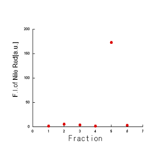
      <a name="Flotation_assay_of_liposome_and_DNA_origami">2-2. Floatation assay of liposome and Rectangular tile(DNA origami)</a>
      <figure>
        <img src="http://openwetware.org/images/1/1d/640pxflotationassay-Todai.jpg" width=300px height=300px >
	<figcaption> Result of agarose gel electrophoresis of the sample of flotation assay

	The result of 1% agarose gel electrophoresis(100V,30min). In this measurement, the fluorescence of Cy5, which is
integrated into DNA origami(Rect tile[1]) ,is observed. Fraction1 is the liquid in the top layer, and fra ction 5 is in the bottom layer. Fraction 6 is the sample retrieved from precipitation. DNA origami solely was also l oaded on the extreme right lane. </figcaption>
      </figure>
      <figure>
        <img src="http://openwetware.org/images/0/0d/300pxNILGraph-Todai.PNG" width=350px height=350px >
	<figcaption> Fluorescence intensity of the samples of flotation assay(DNA Rect tile +liposome)
Although the size of liposome might change during the flotation assay(data not shown), the intensity of the fluoresc ence of NIL(Nile Red, ex 500nm, em 550~700nm ) suggests the amount of lipid membrane,liposome. The fluorescence spectrum of water was subtracted as background
</figcaption>
</figure>
To confirm the flotation assay, mixed tiles(DNA origami) and liposomes were assayed. Five samples (fraction 1,2,..., 5, from the top) were retrieved from supermetant liquid and a sample(fraction 6) from precipitation by the addition of buffer used in assay. When the sample, tile mixed with liposomes, were assayed, tiles were observed in the top layer. The distribut ion of liposomes is observed by the fluorescence of NIL(Nile Red).
</article>
<article>
      <a name="hybridization_of_Cholesterol_Oligo_with_OCK">hybridization of Cholesterol Oligo with OCK</a>
      <figure>
<img src="http://openwetware.org/images/d/d7/480px_OCKchol-Todai.png" width=480px height=300px >
</figure>
The result of 1 % agarose gel electrophoresis showed that the band of sample 5 and 7 were smeared, showing the successful of hybridization of cholesterol oligo with OCK (Langecker et al. (2012)). We concluded that the optimized condition for hybridization is: 1 hour incubation at room temperature with 5 times excess cholesterol oligo to OCK.
</article>
<a name="Comparision_of_dimerization">3. Comparision of dimerization method</a>
<article>
      <a name="Click_reaction_via_(3+2)_cycloaddition">
      3-1. Click reaction via (3+2) cycloaddition[4]
      </a>
<figure>
        <img src="http://openwetware.org/images/a/ab/450pxclick0828-Todai.jpg" width=480px height=360px >
	<figcaption> Result of urea gel electrophoresis of the sample of click reaction
</figcaption>
</figure>
Copper(Ⅰ) catalyzed click reaction was used to dimerize of oligo DNA(length of 20bp and 14bp) . The time cause of the reaction indicate that the click reaction is so quick(<5min).
<article>
<a name ="Azobenzene">3-2. Azobenzene</a>
<a name="Synthesis_of_Tube(Research_for_azobenzene)"> 3-2-1. Synthesis of tube[2],[3](Resear ch for azobenzene)</a>
<figure>
        <img src="http://openwetware.org/images/b/b2/640x360px_tube_result-Todai.png" width=640px height=360px >
        <figcaption>
        results of the electrophoresis of DNA-tube

        </figcaption>
</figure>
We examined how DNA-tube was synthesized efficiently by using the method which is introduced in “Rapid Folding of DNA into Nanoscale Shapes at Constant Temperature” (Jean-Philippe J. Sobczak et al, Science, 2012, 338, 1458) [2]. In the figure above, two bands derived from scaffold or DNA-tube were showed with cursors. At 56.4℃ , the scaffold band was diminished. In contrast, the DNA-tube band was concentrated. In fact, the ratio of DNA- tube band to scaffold band was the greatest at this temperature, which means we succeeded in synthesizing DNA- tube efficiently and improving the yield of DNA-tube.
<article>
<a name="Synthesis_of_Motif(Research_for_azobenzene)"> 3-2-2. Synthesis of Motif[5]</a>
<figure>
        <img src="http://openwetware.org/images/a/a8/480px_electrophoresis_of_T-motif_improved-Todai.png" width=480px h	eight=360px >
        <figcaption>
         Result of agarose gel electrophoresis of T-motif (wheel)


         The result of 1.5% agarose gel electrophoresis(100V) for 33min.
        </figcaption>
</figure>
In this measurement, it is difficult to distinguish between final structure band and monomer band. Next time, Native PAGE will be used instead of agarose gel.
<article>
<a name="Protocols"> Protocols</a>
<article>
<a name="Assembling_of_OCK">Assembly of OCK [2]</a>
<article>
| M13mp18ss | 4.5 ul |
|---|---|
| Staple mix | 4.5 µL |
| 10x OCK buffer* | 1 µL |
*...10x OCKbuffer(f.100 ul)
| Tris-HCl(ph 7.5) | f.50 mM | 1 M | 5 µL |
|---|---|---|---|
| EDTA-Na(pH 8) | f.10 mM | 0.5 M | 2 µL |
| MgCl2 | f.200 mM | 1 M | 20 µL |
| NaCl | f.500 mM | 5 M | 1 µL |
| MQ | - | - | 72 µL |
- mix the solutions.
- It was annealed at 85 °C for 25 °Cmin and then at 52 °C for 3 or 4 hours.
</article>
<article>
      <a name="Transmission_electron_microscopy">
      Transmission electron microscopy(TEM)
      </a>
The procedure of TEM was refered to previous researches[6].
   </article>


  <article>
2.Flotation assay
<a name="Preparation_of_SUVs">Preparation of SUVs</a>
Type 1: POPC 100%
| 150mM aqueous KCl solution | 3mL |
|---|---|
| POPC | 3mg |
| Chloroform (99.0%) | 3mL |
| 40μM Nile Red solution | 0.1mL |
- POPC were dissolved in 3mL of Chloroform.
- A lipid film was formed by evaporating 3mL of POPC solution in a 50mL eggplant flask, using a rotational evaporator for 5 minutes.
- The flask was kept under vacuum overnight to evaporate remaining chloroform.
- The lipid film was resuspended in 3mL of a 150mM aqueous KCl solution.
- The solution was filtered through 200nm polar filter with extruder to even the size of liposome.
- The size of liposome was measured with DLS (Viscotek 802 DLS).
- The solution was kept at 3 degree C until usage.
</article>
<article>
Type 2: POPC 50%, POPG 50%
| Lipid mix* | 3 ml |
|---|---|
| 150 mM aqueous KCl solution | 1 µL |
*...Lipid mix
| 5 mg/mL POPC | 0.1 mL |
|---|---|
| 5 mg/mL POPG | 0.1 mL |
| 10 uM NileRed solution2 | 0.13 mL |
| Chloroform | 2.67 mL |
- A lipid film was formed by evaporating 3 ml of lipid mix in a 50 ml eggplant flask, using a rotational evaporator (EYELA, model#N1110) for 10 mins.
- The flask was kept under vacuum overnight to evaporate remaining chloroform.
- The lipid film was resuspended in 1 mL of 150 mM aqueous KCl solution.
- Lipid suspended solution was filtered through 100nm polar filter using extruder (Avanti) to prepare uniformly-sized liposome.
- The size of liposome was measured with DLS (Viscotek 802 DLS).
- The solution was kept at 3℃ until usage.
</article>
<article>
<a name="Preparation_of_GUVs">Preparation of GUVs</a>
| Lipid mix* | 3 ml |
|---|---|
| 150 mM aqueous KCl solution | 1 µL |
*...Lipid mix
| 5 mg/mL POPC | 0.1 mL |
|---|---|
| 5 mg/mL POPG | 0.1 mL |
| 10 uM NileRed solution2 | 0.13 mL |
| Chloroform | 2.67 mL |
- A lipid film was formed by evaporating 3 ml of lipid mix in a 50 ml eggplant flask, using a rotational evaporator (EYELA, model#N1110) for 10 mins.
- The flask was kept under vacuum overnight to evaporate remaining chloroform.
- The lipid film was resuspended in 1 mL of 150 mM aqueous KCl solution.
</article>
<article>
<a name="Protocol_Hybridization_of_cholesterol_oligo_with_OCK">Hybridization of cholesterol oligo with OCK</a>
| OCK | 48 µL |
|---|---|
| Cholesterol oligo (0.32, 0.64, 3.2, 6.4 µM) | 100 µL |
Marker; GeneRuler DNA Ladder Mix (Fermentas, GeneRuler DNA Ladder Mix #SM0331)
- Centrifuge for 16 minutes at 100 krpm at 4 ℃ using TLA 100.2 rotor (BECKMAN COULTER) with Ultracentrifuge (BECKMAN COULTER, Optima MAX-XP).
- Each sample was mixed and incubated as shown below:*
*...Table1.
| Sample No. | 1 | 2 | 3 | 4 | 5 | 6 | 7 | 8 |
|---|---|---|---|---|---|---|---|---|
| Purified OCK (40 µM) | 6 µL | 6 µL | 6 µL | 6 µL | 6 µL | 6 µL | 6 µL | 6 µL |
| 0.32 µM Cholesterol oligo | 1.5 µL | 1.5 µL | - | - | - | - | - | - |
| 0.64 µM Cholesterol oligo | - | - | 1.5 µL | 1.5 µL | - | - | - | - |
| 3.2 µM Cholesterol oligo | - | - | - | - | 1.5 µL | 1.5 µL | - | - |
| 6.4 µM Cholesterol oligo | - | - | - | - | - | - | 1.5 µL | 1.5 µL |
| [Cholesterol oligo]/[OCK] (see #Note) | 1/2 | 1/2 | 1 | 1 | 5 | 5 | 10 | 10 |
| Incubation time [min] | 60 | 30 | 60 | 30 | 60 | 30 | 60 | 30 |
- Note; OCK has 4 cholesterol oligo binding sites. Therefore, we devided the molar ratio of cholesterol oligo to OCK with 4.
</article>
  <article>
  <article>
<a name="Flotation_assay_[OCK]">Flotation assay [OCK]</a>
| OCK | 100 µL |
|---|---|
| Cholesterol hybridized OCK | 100 µL |
| Liposome (1 mg/mL SUVs) | 100 µL |
| 2.25 M Sucrose buffer* | 500 µL |
| 1.6 M Sucrose buffer** | 900 µL |
| 150 mM aqueous KCl solution | 100 µL |
| 1×Flotation buffer** | 600 µL |
*...2.25 M Sucrose buffer
| HEPES-KOH (pH 7.6) | 50 mM |
|---|---|
| KCl | 100 mM |
| MgCl2 | 20 mM |
| Sucrose | 2.25 M |
**...1.6 M Sucrose buffer
| HEPES-KOH (pH 7.6) | 50 mM |
|---|---|
| KCl | 100 mM |
| MgCl2 | 20 mM |
| Sucrose | 1.6 M |
***...1×Flotation buffer
| HEPES-KOH (pH 7.6) | 50 mM |
|---|---|
| KCl | 100 mM |
| MgCl2 | 20 mM |
- Each sample was mixed as shown below:****
****...Table1. Breakdown of Samples
| Sample No. | 1 | 2 | 3 | 4 |
|---|---|---|---|---|
| OCK | 50 µL | 50 µL | - | - |
| Cholesterol hybridized OCK | - | - | 50 µL | 50 µL |
| Liposome | 50 µL | - | 50 µL | - |
| 150 mM aqueous KCl solution | - | 50 µL | - | 50 µL |
| 2.25 M Sucrose buffer | 125 µL | 125 µL | 125 µL | 125 µL |
</article>
</article>
<article>
3. Comparision of dimerization method
<article>
<a name="Dimerization_of_OCK--using_biotin,_streptavidin_and_click_ reaction">Dimerization of OCK--using biotin, streptavidin and click reaction</a>
| OCK (90 nM) | 8 µL |
|---|---|
| Streptavidin (190 nM) | 2 µL |
| CuSO4 aq (8 mM) | 1 µL |
| THTA (32.5 mM) | 1 µL |
| Sodium ascorbate (3.25 mM) | 1 µL |
- 7.4 µL of OCK and 1 µL Streptavidin (190 nM) were mixed and kept at room temperature (27 ℃) for an hour. (Mix1)
- 10 µL of Mix1 and 1 µL of Sodium ascorbate (3.25 mM) were mixed and then 1 µL of CuSO4 aq (8 mM) was added into that solution.
- The solution was mixed and 1µL of THTA (20 mM) was added in it and mixed.
- That solution was kept at room temperature (27 ℃) for a day.
</article>
      <a name="Click_reaction_via_(3+2)_cycloaddition">
      Click reaction via (3+2) cycloaddition
      </a>
| azide solution (10μM) | 3μL |
|---|---|
| alkyne solution (10μM) | 3μL |
| CuSO4 solution (50mM) | 1μL |
| THTA solution (100mM) | 1μL |
| sodium ascorbate solution (100mM) | 1μL |
- The above all solutions were mixed, using a vortex.
- The solution was kept at room temperature.
   </article>


  <article>
<a name="Accelerated_Click_reaction">Accelerated Click reaction (using streptavidin to make the aklyne and azide reactive groups close) </a>
| 2x barrel buffer | 6 µL |
|---|---|
| alkyne oligo (carrying biotin) (15 µM) | 1 µL |
| azide oligo (carrying biotin) (15 µM) | 1 µL |
| streptavidin (500 µM) | 1 µL |
- mix reagents
- incubate the tube at 37 ℃ for indicated reaction time.
- boil at 95 ℃ for 30 minutes to break down streptavidin
</article>
<article>
<a name="Click_reaction_(using_hybridization_to_make_the_aklyne_and_azide_reactive_groups close)">Click reaction (using hybridization to make the aklyne and azide reactive groups close) </a>
| 2x barrel buffer | 7 µL |
|---|---|
| alkyne oligo (15 µM) | 1 µL |
| azide oligo (15 µM) | 1 µL |
| scaffold (15 µM) | 1 µL |
- mix reagents
- incubate the tube at 37 ℃.
- add loading buffer into the reaction mixture and boil at 95 ℃ for 5 minutes to denature the double strand to single strand.
</article>
<a name="Click_reaction_(copper_catalyst-free)">Click reaction (copper catalyst-free)</a>
| 2x barrel buffer | 7 µL |
|---|---|
| alkyne oligo (15 µM) | 1 µL |
| azide oligo (15 µM) | 1 µL |
| scaffold (15 µM) | 1 µL |
| 1M Tris (pH 7.5) | 5 µL |
|---|---|
| 0.5M EDTA | 2 µL |
| 5M NaCl | 1 µL |
| MQ | 32 µL |
- mix reagents
- incubate the tube at 37 ℃.
- add loading buffer into the reaction mixture and boil at 95 ℃ for 5 minutes to denature the double strand to single strand.
</article>
<article>
      <a name="Research_for_azobenzene)">
      3-2. Research for azobenzene
      </a>
The procedure of synthesis of tubes and motifs were refered to previous researches([2],[3],[5])
</article>
<a name="Reference"> Reference</a>
       <a name="proref-1">
       [1] Folding DNA to create nanoscale shapes and patterns
       </a>
Nature 440, 297–302 (2006)
       <a name="proref-1">
       [2] Rapid Folding of DNA into Nanoscale Shapes at Constant Temperature
       </a>
Science, 2012, 338, 1458
       <a name="proref-1">
       [3] Transcription Regulation System Mediated by Mechanical Operation of a DNA      &nbs	p;Nanostructure
       </a>
Journal of the American Chemical Society, 2012, 134, 2852-2855
       <a name="proref-1">
       [4] the protocol of Jena Bioscience GmbH
       </a>
          <a target="_blank" href="http://www.jenabioscience.com" style="color:#e00000">
          http://www.jenabioscience.com</a>
       <a name="proref-1">
       [5] Substrate-Assisted Assembly of Interconnected Single-Duplex DNA Nanostructures
       </a>
Angewandte Chemie International Edition,2009,48(37),6820–6823
       <a name="proref-1">
       [6] A primer to scaffolded DNA origami.</a>
Nat Methods 221-229 (2011, Mar;8(3))
 </article>


  <footer style="position:relative;left:400px">

   Copyright © Todai nanORFEVRE, all rights reserved.

  </footer>
</body> </html>
