Biomod/2014/Kashiwa/Experiments: Difference between revisions
No edit summary |
No edit summary |
||
| Line 185: | Line 185: | ||
<br><br><br> | <br><br><br> | ||
<div class="left"><img src="http://openwetware.org/images/7/7d/Emission_of_ini.jpg"><img src="http://openwetware.org/images/ | <div class="left"><img src="http://openwetware.org/images/7/7d/Emission_of_ini.jpg"><img src="http://openwetware.org/images/c/c2/Emission_of_ini_20%25.jpg"></div> | ||
<p class="ex">グラフを作った。考察。</p> | <p class="ex">グラフを作った。考察。</p> | ||
Revision as of 14:06, 29 August 2014
<html> <head>
<script type="text/javascript" src="http://code.jquery.com/jquery-1.9.1.min.js"></script>
<script type="text/javascript"> new Image(120,80) = "http://openwetware.org/images/7/7a/Logo2Kashiwa.png";
new image(120,80) = "http://openwetware.org/images/1/1d/Logo2.5.png";
new image(71,143) = "http://openwetware.org/images/2/2a/Top1.gif";
new image(71,143) = "http://openwetware.org/images/f/f0/Top2.gif";
new image() = "http://openwetware.org/images/3/38/BackgroundKashiwa.png";
new image() = "http://openwetware.org/images/9/9c/BackgroundKashiwa2.png"; </script>
<script type="text/javascript">
  $(function(){
     $("#menu li").hover(function(){
        $("ul",this).show();
     },
     function(){
        $("ul",this).hide();
     });
  });
</script>
<script type="text/javascript">
</script>
<META http-equiv="Content-Style-Type" content="text/css">
<style type="text/css">
.page-back {
position: fixed;
bottom: 25px;
right: 25px;}
p.paragraph {
 font-size :100%;
 line-height: 135%;
 margin: 10px 25px;
}
p.reference {
 font-size :90%;
 line-height: 135%;
 margin: 10px 20px;
}
p.indent-paragraph {
 font-size :110%;
 line-height:1.5;
 margin:0 30px;
 text-indent: 1em;
 }
h1.title a{
 font-size :100%;
 display: block;
 text-decoration: none;
 color: #000000;
font-weight:bolder;
 border-left: solid 5px #e00000;
 }
h2.reference a{
 font-size :90%;
 display: block;
 text-decoration: none;
 border-left: solid 5px;
 }
body {
 font-size: 12px;
 font-family: Frutiger, Helvetica, Arial;
 background-color: #B5D3FF;
 overflow-y:scroll;
 overflow-x:hidden;
} article {
background-color: #ffffff
} .container {
  background-color: #ffffff;
  margin-top:0px
}
.OWWNBcpCurrentDateFilled {display: none;}
}
- column-content
{
  width: 0px;
  margin: 0;
  padding: 0;
  float:left;
} .firstHeading {
  display:none;
  width:0px;
}
- globalWrapper
{
  width:900px;
  background-color: #ffffff;
  margin-left: auto;
  margin-right: auto;
}
- column-one
{
  display:none;
  width:0px;
  background-color: #ffffff;
}
- content
{
  margin: 0px;
  align: center;
  padding: 0px 12px;
  width:876px;
  border: 0;
}
- bodyContent
{
  width: 800px;
  padding: 0px 38px;
  align: center;
  background-color: #ffffff;
  position:relative;
}
- column-content
{
  width: 900px;
  background-color: #ffffff;
}
- footer
{
  position: center;
  width: 900px;
}
div.menubar {
  position: fixed;
  background-color: none; /* バーの背景色 */
  opacity: 0.95;
  border-top: 0px double white; /* バーの上端線 */
  border-bottom: 0px double white; /* バーの下端線 */
  min-width: 900px; /* メインメニュー全部が収まる最低横幅 */
  z-index:3
}
div.menubar ul#menu {
  margin: -33px 0px 0px -50px; /* メニューバー外側の余白 */
  padding: 0px; /* メニューバー内側の余白 */
  height: 80px; /* メニューバーの高さ */
  list-style-type: none;
}
div.menubar ul#menu li {
  min-width: 110px; /* メニュー項目の横幅 */
  height: 80px; /* メニュー項目の高さ(「メニューバーの高さ」と一致させる) */
  float: left;
  list-style-type: none;
  list-style-image: none;
  position: relative;
}
div.menubar ul#menu a {
  text-decoration: none; /* メニュー項目の装飾(下線を消す) */
  display: block;
  background-color: #1F003E;/* メニュー項目の背景色 */
  color: white; /* メニュー項目の文字色 */
  line-height: 80px; /* メニュー項目のリンクの高さ(「メニュー項目の高さ」と一致させる) */
  text-align: center; /* メインメニューの文字列の配置(中央寄せ) */
  width: 100%;
  height: 100%;
}
div.menubar ul#menu a:hover {
  background-image: url("http://openwetware.org/images/3/38/BackgroundKashiwa.png"); /* メニュー項目にマウスが載ったときの背景色 */
  color: #8B008B; /* メニュー項目にマウスが載ったときの文字色 */
}
div.menubar ul#menu a:active {
  background-image: url("http://openwetware.org/images/3/33/Background2.png"); /* メニュー項目にマウスが載ったときの背景色 */
  color: #8B008B; /* メニュー項目をクリックした時の文字色 */
}
/* メニューバー直後のClearfix */ div.menubar ul#menu {
zoom:1;
} div.menubar ul#menu:after {
height: 0; visibility: hidden; content: "."; display: block; clear: both;
}
div.menubar ul#menu ul.sub {
  background-color: #1F003E; /* サブメニュー全体の背景色 */
  border-width: 0px 0px 0px 0px; /* サブメニュー全体の枠線の太さ */
  border-style: solid; /* サブメニュー全体の枠線の線種 */
  border-color: #191970; /* サブメニュー全体の枠線の色 */
  margin: 0px;
  padding: 0px;
  display: none;
  position: absolute;
}
div.menubar ul#menu ul.sub li {
  width: 110px;
  height: 50px; /* サブメニュー1項目の高さ */
  border-width: 0px 0px 0px 0px; /* サブメニュー1項目の枠線の太さ */
  border-style: solid; /* サブメニュー1項目の枠線の線種 */
  border-color: #191970; /* サブメニュー1項目の枠線の色 */
  z-index: 3
}
div.menubar ul#menu ul.sub li a {
  line-height: 50px; /* サブメニュー1項目の行の高さ(「サブメニュー1項目の高さ」と合わせる) */
  text-align: center;  /* サブメニュー1項目の項目名の配置(中央寄せ) */
  text-indent: 0px;  /* サブメニュー1項目の項目名前方の余白 */
}
div.menubar ul#menu ul.sub li a:hover {
  background-image: ('back');
  background-size: 100% auto;
  background-color: #FF00FF; /* サブメニュー項目にマウスが載ったときの背景色 */
  color:  #8B008B; /* サブメニュー項目にマウスが載ったときの文字色 */
}
div.menubar ul#menu ul.sub li a:active {
  background-image: ('backclick') /* メニュー項目にマウスが載ったときの背景色 */
  backround-size: 100% auto;
  color: #8B008B; /* メニュー項目をクリックした時の文字色 */
}
</style>
</head>
</html>
<html>
<head> <style>
div#subContents {
   width: 200px;
   background: #FFFFFF;
   position: fixed;
   top: 100px;
   left: 15px;
   z-index: 20;
}
- html div#subContents {
   position: absolute;
   top: expression(eval(document.documentElement.scrollTop+100));
}
.category {
  border-style: double;
  border-width: thick;
  border-color: #483D8B;
  font-size: 12px;
  list-style-type:none;
  list-style-image:none;
  padding: 10px 3px 15px 8px;
  margin: 0px;  }
h1.big{
 font-size :24px;
 display: block;
 text-decoration: none;
 color: #000000;
font-weight:bolder;
 margin: 25px 35px;
 }
h1.small{
 font-size :20px;
 display: block;
 text-decoration: none;
 color: #000000;
font-weight:bolder;
}
div.name{
 font-size :16px;
 color: #191970;
 font-weight:bolder;
 }
p.ex{
 font-size :100%;
 margin: 10px 65px;
 }
div.left img { float: left; position: relative; margin: 10px 5px 10px 65px; width: 300px; height: 213px; }
div.right img { position: relative; margin: 10px 65px 10px auto; width: 300px; height: 213px; float: right; }
div.tateleft img { float: left; margin: 0px 5px 0px 65px; height: 300px; }
div.tateright img { float: right; margin: 0px 65px 0px 5px: height: 300px; }
</style>
- <a href="#1">
</a>
Experiments
- The Receptor
- <a href="#1-1" title="CSS リファレンス一覧">Folding the Receptor</a>
- <a href="#1-2" title="Webデザインの一覧">Forming a Receptor dimer</a>
- <a href="#1-3" title="CSS ビギナー(初心者)向け一覧">Emitting the Initiator</a>
- <a href="#1-4" title="Photoshopの一覧">Embedding on the liposome</a>
- The Motor
- <a href="#2-1" title="(X)HTMLの一覧">Folding the Motor-Monomer</a>
- <a href="#2-2" title="Web関連情報の一覧">Forming the Motor-Polymer</a>
</head>
<body>
<a name="1"></a>
EXPERIMENTS
|
The Receptor |
<a name="background"> Direction</a>
We planned to do experiments in several steps. First, we design the Receptor. Then, the following experiments are conducted in parallel.
• Form the Receptor dimer by using thrombin.
• Emit the Polymerization Initiator by demerization.
• Embedding of Receptor on the liposome.
| <img src="http://openwetware.org/images/6/62/Experimentrece1.jpg" width="250px"> | <img src="http://openwetware.org/images/f/f4/Receptorstep2.jpg" width="250px"> | <img src="http://openwetware.org/images/4/4a/Experimentrece3.jpg" width="250px"> |
<a name="1-1"></a>
Finally, after success in the experiments, we combine them to attain the dimerization and the Initiator emission on the liposome.
<a name="background"> Experiments</a>
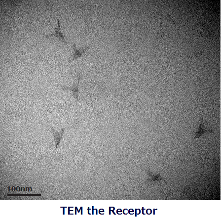
Folding the Receptor
Folding of the Receptor was confirmed by TEM and 1% agarose gel electrophoresis analysis.
The folded structure of the Receptor was clearly appeared in the TEM image.
<a name="1-2"> </a>
Forming the Receptor dimer
In this experiment, the dissociation constant (Kd) of thrombin and thrombin-binding aptamers was estimated. The purpose is to confirm the binding ability of each Aptamer (Aptamer A and B) with a thrombin before checking dimerization of the Receptor hetero-units. Binding of the aptamers with thrombin was analyzed with Native-PAGE.
We could confirm that aptamers bind to thrombin based on these two results. First, in the figure 1, there is a band in addition to aptamer band in the lane #? (aptamer + x10 (or x 20) trhombin). Second, in fig.2, the bands of aptamers are weaker in the lanes (added thrombin 50 or 25 times as much as aptamers) than the other lanes, and we attributed these results to the formation of the complexes of thrombin and aptamers. In addition, the Kd of aptamers and thrombin is estimated about 100~500nM by measuring the intensity of band with the software (Image J).
<a name="1-3"> </a>
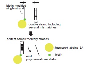
Emitting the Initiator
実験の説明、ゲル電気泳動結果について
グラフを作った。考察。
<a name="1-4"> </a>
Embedding on the liposome
In order to certify the penetration of the Receptors to liposomes, a flotation assay was conducted.
In preparation for a floation assay, cholesterol oligomers were hybridized to the Receptors for penetration. The hybridization was confirmed by 1% agarose gel electrophoresis analysis.
In flotation assay, we divided mixture with the Receptors and the liposomes to a few fractions, each of them was analyzed by fluorescence spectrophotometer and 1% agarose gel electrophoresis. This assay showed the distribution of liposomes and the Receptors, therefore we can distinguish the Receptor embedded into the liposomes from the free Receptors floating in solution.
The fluorescence intensity of NileRed in each fraction (NileRed dyes the membrane of liposomes.)
The result of fluorescence spectrophotometer (JASCO, FP-6500) showed that liposome distributed mostly in fraction 3.
The ratios of the Receptor in each fraction were analyzed by the density of band.
In sample1 (cholesterol + / liposome +), the ratio of the Receptor in fraction3 was a bit larger than that in fraction2. In contrast, in sample 2, 3 and 4, the ratio of the Receptor in fraction3 was smaller than that in fraction2. As liposomes were mostly seen in fraction3 in both sample 1 and 3, and the ratio of the Receptor in each fraction was different only in sample1, we concluded that this difference was caused by penetration of the Receptor to liposomes.
<a name="background"> Next Steps</a>
In these experiments, we achieved to construct the Receptors and insert them into the liposomes. The very basics of others, formation of the Receptor dimers and emission of the Initiator, were also confirmed. Therefore, we are planning to approach the ones following for next steps.
• Confirm the more advanced mechanism of forming the Receptor dimers and emitting the Initiator.
• Examine conditions for dimerization of the Receptor heterounits on the liposomes, may need changes in design of the Receptor.
<a name="2"> </a>
|
The Motor Monomer |
<a name="background"> Direction</a>
We planned to do experiments in several steps. First, we design the Motor-Monomer and assay the formation of a simple Motor-Polymer. Then, the following experiments are conducted in parallel.
• Control the initiation of the polymerization (forming ring structures and ring-opening polymerization).
• Put the Motor-Monomers into the liposome.
| <img src="http://openwetware.org/images/4/4e/Experimentmonomer1.jpg" width="300px"> | <img src="http://openwetware.org/images/e/ef/Experimentmonomer2.jpg" width="300px"> |
<a name="2-1"></a>
Finally, after success in the experiments, we combine them to attain the polymerization in the liposome.
<a name="background"> Experiments</a>
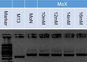
Folding the Motor-Monomer
<img src="http://openwetware.org/images/b/b6/Monomerjouken.png" align="right" style="padding-right:65px">
The assembly condition of the Motor-Monomer was optimezed with concentration of MgCl2, annealing temperature and time. The resulting structure was analyzed by agarose gel electrophoresis.
Multimers (e.g. dimmers) were appeared at lower temperature, so optimum temperature of annealing was 55.0℃.
(Annealing temperature : 55.0℃, M13 : staples = 1 : 2)
The band of monomer at 16mM is bending and its migration distance is different from the one of MoN. It shows that the Monomers were not annealed correctly at 16 mM. There seemed to be little difference at other concentration, so optimum concentration of MgCl2 was the range 10 to 14mM.
(Annealing temperature : 55.0℃, MgCl2 : 10mM)
At the ratio of 6 and 8 a band of a monomer is weak and migrating distance was shorter than the one of monomer. As the graph showed, the ratio of dimer to monomer was low in lane 1 and 2, so optimum ratio of staples to scaffold was 2 or 4.
<a name="2-2"></a>
Folding of the Motor-Monomer was corroborated by PAGE analysis. The folding, however, was not verified by TEM. We considered it was because of hollow structures of the Motor-Monomers.
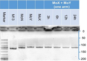
Forming the Motor-Polymer
In this experiment, the effect of arm number on polymerization was examined. Two samples, one has three arms extruded from the body and the other has one, was compared. The Monomers comprise two similar structures, the Motor-Monomer A and the Motor-Monomer B. The polymerization was confirmed by mixing the Monomer A and the Monomer B.
The ratio of dimers of three arms monomers roughly increased with time, while the ratio did not change significantly for one arm monomers. It confirmed the polymerization of the Monomers with three arms.
The Monomer concentration was optimized.
The ratio of dimers was high at high monomer concentration of three arms monomers, while for one arm monomers, the ratio was not different from that of monomer only condition. It meant Polymerization of three arms monomers proceeds efficiently at high concentration of monomers in solution.
<a name="background"> Next Steps</a>
Reference
</body>
<footer style="position:relative; left:600px;"> © 2014 UTokyo Chem & Bio </footer>
</html>
