IGEM:Harvard/2006/DNA nanostructures/Notebook/2006-8-23
From OpenWetWare
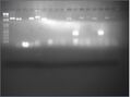
Verify folding of design 5 and 6
Ran gel to verify folding from earlier this week. Also comparing new scaffold with old scaffold.

| Lane | Contents |
| 1 | ladder |
| 2 | p7308 old |
| 3 | p7308 new |
| 4 | c5.0.A (old scaffold) |
| 5 | c5.0.A (new scaffold) |
| 6 | c5.0.C |
| 7 | c5.0.D |
| 8 | c6.0.A |
| 9 | c6.0.B |
| 10 | c6.0.C |
PEG fractionation (from 8-20)
Also including 8% and 10% PEG fractionation for c6.0.A, c6.0.B, and c6.0.C
Streptavidin Depletion Assay
Plans
- Possible Issues and Solutions
- Too much DNA; even if a reasonable amount (say 70% of the biotinylated sites) are binding strep, there wouldn't be a noticeable difference between the bead-treated boxes and non-bead-treated boxes.
- Lower amount of DNA used to half a reaction.
- But the bands aren't that bright as it is, despite the iris being completely open, manual exposure, and use of 5uL of EtBr - possibly due to DNA leeching out during the long (>2hour) electrophoresis? Is that likely at all? But at the same time, my gels have been coming out fainter and fainter as I run longer each time, and yesterday it wasn't even left in buffer for any amount of time at all.
- Run at higher voltage (100V), shorter time (1.5hr) - the difficulty in differentiating the folded from the scaffold might be due recently to the switchover to 0.5xTBE. Take a risk with the higher voltage - check on the gel box every 20 minutes to make sure no boiling is occuring, and scrape off the electrode wire each time.
- But the bands aren't that bright as it is, despite the iris being completely open, manual exposure, and use of 5uL of EtBr - possibly due to DNA leeching out during the long (>2hour) electrophoresis? Is that likely at all? But at the same time, my gels have been coming out fainter and fainter as I run longer each time, and yesterday it wasn't even left in buffer for any amount of time at all.
- Lower amount of DNA used to half a reaction.
- Too little free streptavidin
- Overload with free streptavidin - add from the original stock tube, which is likely safer (in more accurate-for-streptavidin-efficacy) buffer than the dilutions that have been made. This should push the equilibrium towards more complete binding of sites.
- Biotinylated stuff not binding.
- Check by running a control of biotinylated oligos (untreated, beaded, free streped, free then beaded) - there are large amounts of the original tube oligos, so run 40pmol of biotinylated sites (10uM biotinylated sites in the pre-working stock = 10pmoles/uL), or 5uL (to account for inefficient binding and pipetting error) in one lane and 40uL in another, just to make sure that the binding actually works.
- If oligos don't show up in the untreated lane, try SYBR gold staining - ssDNA will show up better at low concentrations that way.
- Check by running a control of biotinylated oligos (untreated, beaded, free streped, free then beaded) - there are large amounts of the original tube oligos, so run 40pmol of biotinylated sites (10uM biotinylated sites in the pre-working stock = 10pmoles/uL), or 5uL (to account for inefficient binding and pipetting error) in one lane and 40uL in another, just to make sure that the binding actually works.
- Suspicion that the magnetic beads may work better because there's no worry of accidentally loading part of the pellet and, thus, bead-bound material.
- Use magnetic beads, concentrated with the MagnaRack initially, and run high concentrations (ie. low total volumes).
- Too much DNA; even if a reasonable amount (say 70% of the biotinylated sites) are binding strep, there wouldn't be a noticeable difference between the bead-treated boxes and non-bead-treated boxes.
Folded:
- 4 rxns each of:
- c5.0.A lidless
- c5.0.Eb
- (4 rxns of c5.0.Fb remain from Katie's foldings on Monday)
- NB: Noticed that there were errors in the pre-working stock table. See c5.0 Table of Working Stocks.
- Had to mix:
- c5.0.Eb
- c5.0.Fb
- Had to mix:
Strep-Deplet Assay Protocol
Test Solutions
 TEST SOLUTIONS
 --------------
 a) c5.0.A lidless (barrel) - 10uL
 b) c5.0.9b (biotinylated oligos - inside) - 10uL
 c) c5.0.Eb (outside biotinylated barrels) - 5uL + 5uL H2O
 d) c5.0.Fb (inside biotinylated barrels) - 10uL (folded 8.22.06)
Protocol
 TEST CONDITIONS
 ---------------
 1. Untreated
 2. Beaded
     - pellet beads and remove initial solution they were packaged in
     - mix with each test solution
     - incubate 5 min
     - add 1x 30mM folding buffer to 40uL (ie. 30uL)
 3. Free-streptavidined
     - test solution in tube
     - mix with 3uL of 1mg/mL stock tube of streptavidin (from NEB)
     - incubate 5min
 4. Free-strep, then beads
     - all of (3)'s steps
     - mix with pelleted beads that had had their initial suspension solutions removed
     - incubate 5 min
Gel
- Loaded 10uL of 1.Untreated and ~38uL of the other test conditions into wells of 2% agarose gel, 5uL EtBr, 0.5x TBE, 10mM MgCl2, 2uL of loading dye each. Ran @ 100V in 0.5x TBE, 10mM MgCl2 for 1hr.
| Lane | Component | Test Condition | Amount |
| 1 | 1kb+ ladder | - | 10uL |
| 2 | p7308 (~42nM) | - | 9uL |
| 3 | barrel (lidless) | Untreated | 10uL |
| 4 | biotinylated oligos | Untreated | 10uL |
| 5 | outside biotinylated barrel | Untreated | 10uL |
| 6 | inside biotinylated barrel | Untreated | 10uL |
| 7 | barrel (lidless) | Beaded | ~38uL |
| 8 | biotinylated oligos | Beaded | ~38uL |
| 9 | outside biotinylated barrel | Beaded | ~38uL |
| 10 | inside biotinylated barrel | Beaded | ~38uL |
| 11 | barrel (lidless) | Free-strep | ~38uL |
| 12 | biotinylated oligos | Free-strep | ~38uL |
| 13 | outside biotinylated barrel | Free-strep | ~38uL |
| 14 | inside biotinylated barrel | Free-strep | ~38uL |
| 15 | barrel (lidless) | Free, bead | ~38uL |
| 16 | biotinylated oligos | Free, bead | ~38uL |
| 17 | outside biotinylated barrel | Free, bead | ~38uL |
| 18 | inside biotinylated barrel | Free, bead | ~38uL |
-
 Brighter Bands
 -
Darker background (doesn't highlight the strange bright front midway up as much)

