20.109(S14):DNA cloning (Day4): Difference between revisions
| Line 121: | Line 121: | ||
#Pick up an ethanol jar, spreader, lighter, and alcohol burner when you are almost ready to begin. Don't forget to wear your safety glasses! | #Pick up an ethanol jar, spreader, lighter, and alcohol burner when you are almost ready to begin. Don't forget to wear your safety glasses! | ||
#Plate 100 μL of each transformation mix onto an LB- | #Plate 100 μL of each transformation mix onto an LB-Kanl plate. After dipping the glass spreader in the ethanol jar, you should pass it through the flame of the alcohol burner '''just long enough to ignite the ethanol'''. '''After letting the ethanol burn off, the spreader may still be very hot''', and it is advisable to tap it gently on a portion of the agar plate without cells in order to equilibrate it with the agar (if it sizzles, it's way too hot). Once the plates are ready, place them in the 37°C incubator overnight; be sure that each is labeled with your team color, gull sample ID, and initials. One of the teaching faculty will remove them from the incubator and set up liquid cultures for you to use next time. | ||
==For next time== | ==For next time== | ||
Revision as of 09:19, 3 January 2014
Introduction
Welcome back! Last time you performed PCR on the DNA pool you extracted from a gull sample. Specifically, you used primers designed to amplify bacterial 16S rRNA gene fragments from this complex pool. Now you want to systematically analyze individual fragments from this 16S DNA pool, which is aided by a process called cloning.
In traditional cloning, an insert is ligated to a backbone to create a circular piece of DNA called a vector. Typically, the insert and backbone are cut with special proteins called restriction enzymes (more about those in Module 2!) that yield complementary overhangs at the ends of the two pieces of DNA. The enzyme DNA ligase then chemically bonds the insert and backbone together by catalyzing formation of a phosphodiester bond. Ligated vectors are selected in bacteria by including an antibiotic resistance marker on the backbone, and growing the bacteria in said antibiotic. (We’ll say more about that procedure below.) This method is especially suitable for cloning with the purpose of creating a well-defined piece of DNA.

When diverse products must be cloned for subsequent analysis, as in our case, we would like to omit some steps from the above workflow. A few methods exist for cloning PCR products directly. The most popular approach is dubbed TA cloning, and it exploits the fact that polymerases such as Taq add an adenine to the 3’ end of PCR products. This single-base overhang alone can be used in a process very much like traditional cloning. Recall, however, that we used Pfu polymerase precisely for its precision, to improve our chance of successful amplification from a “difficult” sample such as stool. Pfu has proofreading capabilities, meaning that it doesn’t add spurious adenines.
Pieces of DNA that do not share complementary overhangs, that indeed have no overhangs at all but instead have blunt ends, can also be ligated together. There are several mechanisms by which this ligation may be accomplished…. One is blah. We will simply do bleu.
Here the biology of DNA topoisomerase I is exploited. The backbone includes a CCCTT sequence, which is cut by the topoisomerase, resulting in so-called two vector arms. The vector arms remain bound to the topoisomerase, and can ligate back to each other or to a newly introduced insert sequence. The reaction mixture – buffer, vector arms, and insert (in our case the PCR from M1D3) – is then added to bacteria.

ADD BACK SOME DETAILS
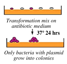
Bacteria can take up foreign DNA in a process called transformation, during which a single plasmid enters a bacterium and, once inside, replicates and expresses the genes it encodes. Most bacteria do not exist in a transformation-ready state, but can be made permeable to foreign DNA -- by chemical or other means -- and are then termed competent. After transformation, the bacteria are plated on an antibiotic-containing agar medium. In your case, the vector arms have an ampicillin resistance gene, and thus only bacteria that took up the vector can grow on ampicillin-containing plates, while untransformed cells will die before they can form a colony. Notice that a vector-containing bacterium will grow whether or not that vector contains a PCR product insert.
We would rather not waste our time analyzing colonies that contain pure vector DNA! Thus, the reaction mixture is added to a particular strain of bacteria that has a mutation in…
The cloning process as a whole is summarized in the figure below.

So today you’ll ligate and transform your PCR product pool, but only after visualizing that everything looks as expected on an agarose gel. Next time you will isolate DNA clones from individual colonies and send them off for sequencing. Then we’ll be just one step away from knowing what bacteria (a sampling of them, in any case!) are present in each gull sample.
Protocols
Part 1: Gel electrophoresis of PCR products
You will use a 1.5% agarose gel to run your three PCRs from last time, as well as a reference lane of molecular weight markers (also called a DNA ladder).
- Add 4 μL of loading dye to each of three eppendorf tubes. No need to change tips in between.
- Loading dye contains xylene cyanol as a tracking dye to follow the progress of the electrophoresis (so you don’t run the smallest fragments off the end of your gel!) as well as glycerol to help the samples sink into the well.
- Now add 20 μL of each reaction from last time (your sample, your partner's sample, and the no template control) to individual eppendorf tubes and label them.
- Flick the eppendorf tubes to mix the contents, then quick spin them in the microfuge to bring the contents of the tubes to the bottom.
- Remember: to quick-spin, hold down the "short" button on your centrifuge for 3-5 seconds, then release.
- Load the gel according to the table below. Up to 4 groups will share each gel: 2 groups per lane.
- To load your samples, draw 20 μL into the tip of your P20. Lower the tip below the surface of the buffer and directly over the well. You risk puncturing the bottom of the well if you lower the tip too far into the well itself (puncturing well = bad!). Slowly expel your sample into the well. Do not release the pipet plunger until after you have removed the tip from the gel box (or you'll draw your sample back into the tip!).
- Once all the samples have been loaded, we will attach the gel box to the power supply and run at 110 V for 45 minutes.
- Later you will be shown how to photograph your gel and, if necessary, to excise the relevant band of DNA. Begin by anticipating where you expect to see your sample band relative to the bands of the DNA ladder, described here.

| Lane | Sample (20 μL) | Lane | Sample (20 μL) |
|---|---|---|---|
| 1 | DNA ladder (load 10 μL) | 6 | DNA ladder (load 10 μL) |
| 2 | Group 1, NTC | 7 | Group 2, NTC |
| 3 | Group 1, sample A | 8 | Group 2, sample A |
| 4 | Group 1, sample B | 9 | Group 2, sample B |
| 5 | BLANK | 10 | BLANK |
Part 1B: OPTIONAL -- Purify 16S PCR band from gel
In many types of cloning, PCR products are purified prior to ligation. For the cloning approach that we are taking, the manufacturer recommends using PCR products directly. Indeed, in pilot experiments, we found that cloning was at least as successful with direct PCR products as with gel-purified PCR products. However, if your PCR resulted in multiple bright bands on your agarose gel, then you will need to isolate the 16S band before proceeding.
In gel purification, a DNA band is melted, then isolated on a silica (SiO2) column similar to the one you used last time. Salt concentration and pH effects, along with ethanol precipitation, will alternately allow for binding and eluting the DNA while washing away contaminants. The protocol is linked separately here to avoid clutter on this page.
Part 2: Ligate purified product to backbone
Will they each clone this time (pricy) or still choose the brighter band? Unlikely to be different because not distributed across two groups this time… REVISE protocol in any case
- First, choose whichever PCR yielded a brighter band on the gel (assuming no non-specific products in either), and perform one ligation and transformation together. Ultimately, you will each prepare your own plate of cells. Please save both extra PCR reactions and leftover ligation mix today. These will be stored frozen in case of later needs.
- For example, if Shannon and Agi both worked with gull sample 842, and Shannon's band was brighter, then Shannon and Agi would perform the procedure below with Shannon's PCR, but they wouldn't throw away Agi's PCR either.
- Prepare a clearly labeled eppendorf tube for the cloning reaction.
- In the following order, combine 3 μL cloning buffer, 2 μL DNA, and 1 μL of vector mix.
- Incubate at room temperature for 5 min, and then move to ice if you are not yet ready to proceed with the next step. Meanwhile, obtain an aliquot of competent cells from the teaching faculty and let them slowly thaw on ice -- takes about 5 min, in fact. Label the side of the tube (and also the top if you like).
- When both cells and reaction are ready, add 1 μL of the reaction to the cells. Do not pipet up-and-down more than once! Instead, gently mix the cells and DNA by twirling the tube in your fingers.
- Hold the tube near the top, so that the cells at the bottom stay cold.
- Incubate the mixture on ice for 20 min.
- Place the tubes in the 42 °C heat block for exactly 45 seconds, and transfer immediately immediately to ice for 2 min more.
- Add 250 μL of warm LB to each sample; do not pipet to mix.
- Place the tubes on the nutator in the 37 °C incubator, and rock them for about 1 hr.
- You may want to put a colored sticky label on the tube so you can quickly identify it later.
- Shortly after starting your incubation, pick up two LB-Amp plates from the incubator and bring them to the front teaching bench. A member of the teaching faculty will demonstrate how to spread 40 μL of X-gal onto the plates. Return your well-labeled plates (team/day/initials/sampleID/date) to the incubator to warm up further.
Part 3: Prepare tubes for liquid O/N cultures
You will make your teaching faculty very happy if you contribute to their preparatory work.
- Please label 8 large glass test tubes with your team color and sample ID, per sample.
- Mix 25 mL LB with 25 μL of kanamycin.
- Using a serological pipet, aliquot 2.5 mL of LB+Kan per tube. These will be used to set up liquid overnight cultures from your 8 colonies for next time.
Part 4: Transform ligated product into cloning strain
- Pick up an ethanol jar, spreader, lighter, and alcohol burner when you are almost ready to begin. Don't forget to wear your safety glasses!
- Plate 100 μL of each transformation mix onto an LB-Kanl plate. After dipping the glass spreader in the ethanol jar, you should pass it through the flame of the alcohol burner just long enough to ignite the ethanol. After letting the ethanol burn off, the spreader may still be very hot, and it is advisable to tap it gently on a portion of the agar plate without cells in order to equilibrate it with the agar (if it sizzles, it's way too hot). Once the plates are ready, place them in the 37°C incubator overnight; be sure that each is labeled with your team color, gull sample ID, and initials. One of the teaching faculty will remove them from the incubator and set up liquid cultures for you to use next time.
For next time
Reagent list
write something here or not accessible to edit
