Biomod/2011/Harvard/HarvarDNAnos:Designs
<html> <head>
<style>
- column-one { display:none; width:0px;}
.container{background-color: #f5f5f5; margin-top:50px} .OWWNBcpCurrentDateFilled {display: none;}
- content {width: 0px; margin: 0 auto auto 0; padding: 1em 1em 1em 1em; align: center;}
- column-content {width: 0px; float: left; margin: 0 0 0 0;padding: 0;}
.firstHeading {display:none; width:0px;}
- globalWrapper{width:1280px; margin:auto}
body {background: #F0F0F0 !important;}
- column-one {display:none; width:0px;background-color: #f0f0f0;}
- content{border:none;margin: 0 0 0 0; padding: 1em 1em 1em 1em; position: center; width: 800px;background-color: #f0f0f0; }
.container{ width: 800px; margin: auto; background-color: #f0f0f0; text-align:justify; font-family: helvetica, arial, sans-serif; color:#f0f0f0; margin-top:25px; }
- bodyContent{ width: 1267px; align: center; background-color: #f0f0f0;}
- column-content{width: 1280px;background-color: #f0f0f0;}
.firstHeading { display:none;width:0px;background-color: #f0f0f0;}
- header{position: center; width: 800px;background-color: #f0f0f0;}
- footer{position: center; width:1280px;}
</style>
</head> </html>
<html><center><a href='http://openwetware.org/wiki/Biomod/2011/Harvard/HarvarDNAnos'><img src=http://openwetware.org/images/1/1c/Hdnaheader.jpg width=800px></a></center></html>
Rectangular Box Container Design Summary

Andersen's box impressed us with its ability to open and close, but we worried about its robustness and tightness as a container.
- Cryo-EM imaging performed by Andersen revealed that the faces are either bent inward or outward.
- Furthermore, the Andersen box is formed from a one-layer DNA sheet and, as such, is held together by five potentially weak seams.
Therefore, with the help of Wei Sun, we have designed our own box, which we feel stands a much better chance of keeping cargo inside and which is more straightforward to fold and to characterize.
See also: Rectangular Box Results, Rectangular Box Methods
Spherical Container Design Summary

In our search for a robust and elegant design, we were inspired by the origami sphere that Dongran Han demonstrated in his 2011 Science paper "DNA Origami with Complex Curvatures in Three-Dimensional Space". The design principles for an origami sphere (and other 3D origami with complex curvatures) employed by Han are the following (see Figure 1):
- Multi-planar arrangement of parallel double helices with in-plane curvature of helices into rings, and
- Curvature across planes caused by different ring sizes and greater distance between crossovers in larger rings than in smaller rings.
See also: Sphere Results, Sphere Methods
Cargo


With a few container designs in mind, our next goal was to provide them with functionality.
- We decided to use 5 nm gold nanoparticle cargo as a test platform for our ability to capture, contain, and controllably release cargo.
- We decided to use 5 nm gold nanoparticles because the sharp contrast they provide under TEM would help us to classify our results easily.
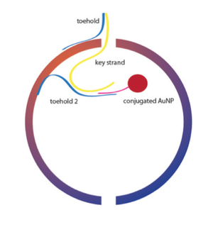
Our primary mechanism for attaching cargo involved conjugating 5 nm gold nanoparticles to ssDNA strands complementary to staple strands extending into the the inside of our containers. Conjugation of gold to ssDNA was made possible by ordering our ssDNA with thiolated 5' or 3' ends. We then designed two processes to release our cargo within our containers: strand displacement and photo-cleavage.
- For the strand displacement method we engineered a staple extension within each structure complementary to a region of the DNA strand conjugated to our gold nanoparticles. The gold nanoparticles would in turn be displaced by a key strand we engineered to be brought into the vicinity of our structure (most specifically for the sphere) by an exterior toehold, and then would bind to a single stranded region of the container's staple extension, and would then proceed by kinetic probability to displace the conjugated AuNP by preferentially binding to its complementary region with the container's staple extension. (See Figure 3 for a depiction of this process in the sphere)
- For the photo-cleavage method, we designed a staple extension into the interior of our containers that contained a region complementary to the DNA bound to our conjugated gold nanoparticles, but also a photo-cleavable spacer prior to the binding site with the gold nanoparticle's DNA strand. Thus, after binding of the AuNP and the subsequent introduction of UV light, the nanoparticle's attachment to the inside of the container would be severed, and the NP would move freely within until subsequent (or simultaneous) opening of the container. (See Figure 4 for a depiction of this process in the sphere.)
See also: Cargo in the Sphere, Cargo in the Box, Nanoparticle Results, Photocleavage Results