Courtney L. Merriam Week 10
Purpose
This journal’s purpose is to work towards the completion of the weekly assignment for week nine. We are meant to examine the amino acid sequences of subjects in order to find the v3 region and compare it to other previously taken DNA sequences using a multiple sequence alignment to find any differences between them.
Methods and Results
- Download the structure file for the paper we read in journal club from the NCBI Structure Database
- The files can be opened with the Cn3D software site. Answer the following:
- Find the N-terminus and C-terminus of each polypeptide tertiary structure.
- Locate all the secondary structure elements. Do these match the predictions made above?
- Yes it seems to match
- Locate the V3 region and figure out the location of the Markham et al. (1998) sequences in the structure.
Work Session for Our Project
- Reselect the subjects for out experiment
- Subjects 13,12,10, and 4 the first and the last visit. Subjects 4 and 10 are the highest progressing subjects and have based on their annual CD4 count. Subject 12 and 13 were the lowest non-progressing subjects based on their annual CD4 Count
- Performed a multiple sequences alignment with all subjects chosen and their clones from first and last visits
- Analyzed data to find a pattern in the amino acid changes
- The Huang paper sequence was compare to the Markham sequences using a protein rendering and analysis program
- The purpose behind this was to find the differences between the Huang paper and the Markham sequences
- Perform a cluster alignment of all the sequences we are using to show the difference between each subject's first and last visit
- The distances were recorded for each subject.
- Create a presentation of the work in progress for next week
Data and Files

- This structure was used to generate the Markham sequences to be compared to experimental subject sequences for comparison.
ClustalDist Results
- Following data is the distance between each subject's first and last visit
- Subject 10: 0.116
- Subject 4: 0.117
- Subject 12: 0.053
- Subject 13: 0.021
Experimental Subjects
Rapid Progressors
- Subject 13-visits 1 and 5
- Subject 12-visits 1 and 8
Non Progressors
- Subject 10-vists 1 and 6
- Subject 4-visits 1 and 4
Amino Acid Mutations Between Subject's First and Last Visits
- Subject 4→ 1 mutation
- Subject 10→ 8 mutations
- Subject 12→ 3 mutations
- Subject 13→ 0 mutations
Conclusion
- Rapid progressors and non progressors were chosen in an even distribution. We hypothesized that there would be a higher number of
amino acid mutations in rapid progressors from the comparison of their first visit to their last. A multiple sequence alignment was done between all involved subjects’ DNA from clones of their first and last visit. The multiple sequence alignment did not show any noticeable pattern of amino acid change between a subjects first and last visit. We then used an amino acid sequence from the Huang article and compared it to the amino acid sequences of our subjects, but there was nothing very different between them.
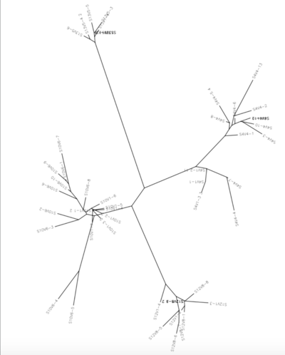
- We then used the Biology Workbench tool ClustalDist to create an unrooted tree that showed the genetic differences in the test subjects. It
also showed a larger difference between the first and last visits of the rapid progressors, where the same was not shown for the nonprogressors, which supports our hypothesis. Still, the analysis of all the mutations across all subjects showed that not all subjects had significant change in amino acids. Subjects 13 and 4 showed minimal amino acid change when first and last visit were compared, and subjects 10 and 12 had significant amino acid mutations. This goes counter to our hypothesis, because the amino acid mutation and lack thereof was equally demonstrated in non-progressors and rapid progressors.
Acknowledgments
I collaborated with Shivum A Desai in class on this assignment. While I worked with the people noted above, this individual journal entry was completed by me and not copied from another source.*Courtney L. Merriam 13:11, 4 November 2016 (EDT):
References
- Kirchherr, J. L., Hamilton, J., Lu, X., Gnanakaran, S., Muldoon, M., Daniels, M., Kasongo, W., Chalwe, V., Mulenga, C., Mwananyanda, L., Musonda, R.M., Yuan, X., Montefiori, D.C., Korber, M.T., Haynes, B.F., & Musonda, R. M. (2011). Identification of amino acid substitutions associated with neutralization phenotype in the human immunodeficiency virus type-1 subtype C gp120. Virology, 409(2), 163-174. DOI: 10.1016/j.virol.2010.09.031
- Markham, R.B., Wang, W.C., Weinstein, A.E., Wang, Z., Munoz, A., Templeton, A., Margolick, J., Vlahov, D., Quinn, T., Farzadegan, H., & Yu, X.F. (1998). Patterns of HIV-1 evolution in individuals with differing rates of CD4 T cell decline. Proc Natl Acad Sci U S A. 95, 12568-12573. dos: 10.1073/pnas.95.21.12568
Useful Links
Clas Page: Bioinformatics Laboratory
| Weekly Assignments | Individual Journal Assignments | Shared Journal Assignments |
|---|---|---|
|
|
|
