Electrophoresis
Electrophoresis Overview
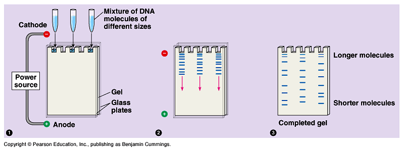
Electrophoresis is a technique primarily used to separate and analyze bands of DNA. In typical biological lab settings, electrophoresis is facilitated through an agarose gel, which possesses channels when cast [1]. Samples of DNA are then injected into each channel, where an electric current run through gel (see Figure 1). The end of the system closer to where the channels are located possesses the cathode head, whereas the oppose end of the system has the anode head. Since nucleic acids (e.g. DNA and RNA) are negatively charged due to their negatively charged backbone, samples travel through the gel towards the positive end of the system, towards the anode [1]. Travelling through the channels allows the DNA samples to separate based on their molecular size, with smaller pieces of DNA travelling further down the channel.
Challenges of Electrophoresis
Since electrophoresis deals with living matter, a prevalent limitation of the technique is contamination. Contamination of either the gel or chemicals (e.g. buffer) used in an electrophoretic experiment would result in contamination of the DNA sample, and thus compromise results [2]. In order to overcome this limitation, it is imperative that electrophoresis be carried out in sterile laboratory settings in order to produce optimal results.
Additionally, electrophoresis is a longer separation process when compared against other microfluidic techniques, which can cause degradation of the working sample [2].
Electrophoresis Innovations and Miniaturization
Miniaturization of electrophoresis has been developed in order to aim for efficiency: maximizing productivity while minimizing area [3]. These innovations to electrophoresis would enable experiments to run simultaneously, as well as reduce the time and cost required for running these experiments [4]. A feature of electrophoresis miniaturization is incorporation of separation channels, which work to increase channel efficiency.
A category of electrophoresis, free-flow electrophoresis (FFE), uses channels with sample continually streaming through them whilst an electric current is applied perpendicular to the channels [5]. Miniaturization of FFE has improved efficiency by enabling analysis of samples in small volumes.
References
[1] Lee, P. Y., Costumbrado, J., Hsu, C. Y., & Kim, Y. H. (2012). Agarose gel electrophoresis for the separation of DNA fragments. JoVE (Journal of Visualized Experiments), (62), e3923-e3923. DOI: http://dx.doi.org/10.3791/3923
[2] Chéry, C. C., Moens, L., Cornelis, R., & Vanhaecke, F. (2006). Capabilities and limitations of gel electrophoresis for elemental speciation: A laboratory's experience. Pure and Applied Chemistry, 78(1), 91-103. DOI: http://dx.doi.org/10.1351/pac200678010091
[3] Pfeiffer, A. J., Mukherjee, T., & Hauan, S. (2004). Design and optimization of compact microscale electrophoretic separation systems. Industrial & engineering chemistry research, 43(14), 3539-3553. DOI: http://dx.doi.org/10.1021/ie034071t
[4] Campana, A. M. G., Baeyens, W. R., Aboul-Enein, H. Y., & Zhang, X. (1998). Miniaturization of capillary electrophoresis systems using micromachining techniques. Journal of Microcolumn Separations, 10(4), 339-356. DOI: http://dx.doi.org/10.1007/978-94-017-2264-3_91
[5] Turgeon, R. T., & Bowser, M. T. (2009). Micro free-flow electrophoresis: theory and applications. Analytical and bioanalytical chemistry, 394(1), 187-198. DOI: http://dx.doi.org/10.1007/s00216-009-2656-5