IGEM:Hong Kong HKUST/Investigations/Effects of ATP concentration to the Ligation Efficiency/Entry Base: Difference between revisions
| Line 85: | Line 85: | ||
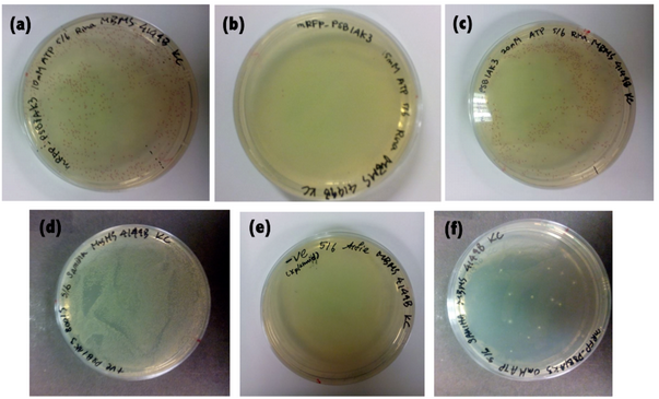
Table 4- Number of red colonies (CFU) on plates containing cells with plasmids ligated at varying concentrations of ATP(10mM, 15mM, 20mM) | Table 4- Number of red colonies (CFU) on plates containing cells with plasmids ligated at varying concentrations of ATP(10mM, 15mM, 20mM) | ||
<br> | <br> | ||
"http://openwetware.org/images/2/2b/Table_4_from_R%26S.PNG" | |||
<br> | <br> | ||
"http://openwetware.org/images/6/60/Plates_from_R%26S.PNG" | "http://openwetware.org/images/6/60/Plates_from_R%26S.PNG" | ||
Revision as of 21:01, 7 June 2015
AuthorsAbstractLigase is an important enzyme involved in joining nicks of DNA strands, especially in forming recombinant DNA. As ATP is required as a co-factor of T4 DNA ligase activity, an experiment was done investigating the effect of ATP concentration on DNA ligation success rate. Two DNA fragments obtained from the BioBrick Parts Registry -BBa_J04450, an mRFP generator, and pSB1AK3, which is a vector with high copy number- were ligated in ligation mixtures with different ATP concentrations. Successful ligations, indicated by the formation of red colonies on gel plate after transformation, which was measured in CFU (Colonies Forming Unit). Due to insufficient sample replicates, the data collected in this experiment is insufficient for suggesting a correlation between the two variables. IntroductionLigation is a process in which two fragments of DNA are joined together in the presence of enzyme ligase. T4 DNA ligase is one of the the enzymes commonly used for ligation in laboratories. It requires ATP as a co-factor to form phosphodiester bonds between the two DNA fragments. In the assembly of genes, optimisation of the ligation process is crucial for obtaining maximal successful DNA constructs. Thus, in this experiment, the effect of ATP concentration on ligation success rate was investigated. 2 plasmids were employed, namely BBa_J04450-pSB3K3 and BBa_B0015- pSB1AK3, to extract mRFP generator and backbone respectively. The ATP concentration was varied by adding different concentration of ATP during ligation of the insert and backbone, i.e. 10mM, 15mM, 20mM. It was hypothesized that the increase of ATP concentration in the ligation mixture increases the ligation efficiency, as ligase requires ATP to carry out ligation. Methods and Material1. Preparation of LB broth 2 bottles of 400mL of LB broth with agar were prepared, each by mixing the below ingredients and made up to 400ml with double distilled water(ddH2O). - Tryptone 4g - Yeast 2g - NaCl 4g - Agar 6g The mixtures were brought to autoclave. One bottle was added with chloramphenicol and the other with ampicillin, then poured into plates and left for solidification. 2. Transformation: After thawing the competent cells in ice, 1 µl of BBa_J04450 in pSB3K3 DNA was added to tubes of competent cells for transformation. A plate of cells without any plasmid was made as negative control. 1ml of LB broth was added to the competent cells and left for incubation. The cells were then grown on kanamycin added LB agar plate overnight with the corresponding antibiotics. 3. Inoculation Two colonies of cells were extracted for inoculation in LB broth overnight. Cells with plasmid BBa_J04450-pSB3K3 were obtained from agar plates from the previous day, and cells with plasmid BBa_B0015- pSB1AK3 was obtained from an agar plates of cells with corresponding plasmids. 4. Miniprep Miniprep was carried out following the standard procedure given in the Mini Plus ™ Plasmid Extraction System Miniprep kit. The DNA concentrations of each sample were obtained by NanoDrop machine. 5. Digestion
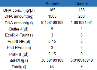
Digestion mixtures following the below recipes were made and incubated at 37 °C for 1 hour. No enzymes were added to the negative controls.
"http://openwetware.org/images/7/74/Table_2_from_R%26S.PNG"
8. Ligation
Insert BBa_J04450 and backbone pSB1AK3 were ligated using the below ligation recipe. Since sample S1(6.5ng/µl) contained the highest concentration of DNA, it was used for the ligation mixture. The ATP concentration was varied from 10mM to 20mM, by adding corresponding volumes of ATP solution(10mM of ATP) to create 15mM and 20mM mixture.
The solution was left to incubate overnight at 16 °C.
Table 3- ligation recipe
"http://openwetware.org/images/e/e2/Table_3_from_R%26S.PNG"
9. Transformation The ligation mixtures after overnight incubation were each transformed into 50 µl of competent cells. The cell mixture was left to incubate for 1 hour at 37 °C to allow sufficient time for recovery. 10. Screening The cells were spread on agar plate with ampicillin resistance and incubated overnight at 37 °C . The number of red colonies formed on each plate were counted the next day. ResultsThe following results were obtained after transforming the ligated products and incubating the transformed cells overnight.
ConclusionThrough data obtained in this experiment, a conclusion for the investigation topic could not be made. This is due to the insufficient sample replicates, which was because of the lack of pSB1AK3 obtained after the purification of gel following gel electrophoresis. In future experiments, more DNA should be used in digestion taking into account that loss of DNA occurs during electrophoresis. A discrepancy of data was observed for 15mM ATP ligation mixture. A significant decrease was observed compared to the 10mM ATP ligation mixture, from 642 to 6 CFU. This result could possibly be due to the following errors: 1) When the 100ul cells were split into half, proper mixing was missed, leading to a non-homogenous cell mixture. The cell solution that was transformed by 15mM ligation mixture had much lower cell number, leading to the low CFU on the agar plate. 2) Although all the transformations were done by the same person, there might be some discrepancies between these transformations, some mistakes or errors affected the 15mM transformation. |