OhioMod2013:Results: Difference between revisions
| (2 intermediate revisions by the same user not shown) | |||
| Line 22: | Line 22: | ||
===Zetasizer=== | ===Zetasizer=== | ||
Determine size and zeta potential of particles. With a high zeta potential we can infer stability, as it can indicate a high repulsion between nanoparticles, hence avoiding flocculation from the emulsion. | Determine size and zeta potential of particles. With a high zeta potential we can infer stability, as it can indicate a high repulsion between nanoparticles, hence avoiding flocculation from the emulsion. | ||
===Fourier-transform Infrared Spectroscopy=== | |||
Confirm citrate functionalization | |||
===Gas Pycnometry=== | ===Gas Pycnometry=== | ||
| Line 33: | Line 35: | ||
===Endurance=== | ===Endurance=== | ||
Store nanoparticles at 25°C and 4°C. Measure rate of precipitation over week. How long can we store the nanoparticles? | Store nanoparticles at 25°C and 4°C. Measure rate of precipitation over week. How long can we store the nanoparticles? | ||
===Uptake Efficiency=== | |||
For liposomal encapsulation efficiency: Use impermeable DNA intercalator dye such as propidium iodide to stain the liposomes before and after ethanol dissolution. Will see an increase in flourescence after ethanol dissolution that correlates with amount of origami in the liposome. | |||
For calcium phosphate encapsulation efficiency: Add DNA intercalator, stain before and after acid dissolution of calcium phosphate. Increase in flourescence correlatees with origami protected by calcium phosphate. | |||
==Serum Assay== | ==Serum Assay== | ||
| Line 42: | Line 49: | ||
==Cytotoxicity== | ==Cytotoxicity== | ||
Perform MTT assay to measure metabolic activity of culture. | Perform MTT assay to measure metabolic activity of culture. Or use brightfield. | ||
==Cellular Uptake== | ==Cellular Uptake== | ||
| Line 60: | Line 67: | ||
Use histoligical staining by Von Kossa method to measure particle uptake into cells. | Use histoligical staining by Von Kossa method to measure particle uptake into cells. | ||
[[Image:VonKossa.png|200px|right|thumb|Von Kossa reaction. Dark cells indicate particle uptake]] | [[Image:VonKossa.png|200px|right|thumb|Von Kossa reaction. Dark cells indicate particle uptake]] | ||
==In vivo== | |||
Use indocyanine green near-infrared imaging to track within body | |||
==Transfection Efficiency== | ==Transfection Efficiency== | ||
Latest revision as of 19:01, 1 July 2013
Results
Nanoparticle Characterizatione
HPLC
Take A260 peaks, determine rate of DNA encapsulation vs free DNA.
Ca:P ratio
Use inductively coupled mass spectroscopy to determine Ca:P ratio.
Zetasizer
Determine size and zeta potential of particles. With a high zeta potential we can infer stability, as it can indicate a high repulsion between nanoparticles, hence avoiding flocculation from the emulsion.
Fourier-transform Infrared Spectroscopy
Confirm citrate functionalization
Gas Pycnometry
Determine density of nanoparticles
TEM
Take direct measurements of ~100 particles using ImageJ software. Determine consistency of size.
Dissolution
Perform titration of nanoparticles until dissolution of nanoparticles. Run gel for DNA origami vs. free DNA after dissolution. Is origami intact?
Endurance
Store nanoparticles at 25°C and 4°C. Measure rate of precipitation over week. How long can we store the nanoparticles?
Uptake Efficiency
For liposomal encapsulation efficiency: Use impermeable DNA intercalator dye such as propidium iodide to stain the liposomes before and after ethanol dissolution. Will see an increase in flourescence after ethanol dissolution that correlates with amount of origami in the liposome.
For calcium phosphate encapsulation efficiency: Add DNA intercalator, stain before and after acid dissolution of calcium phosphate. Increase in flourescence correlatees with origami protected by calcium phosphate.
Serum Assay
Apply DNA encapsulated particles vs free DNA origami vs PEGylated particles to bovine serum. Measure intact origami after period of time via agarose gel electrophoresis.
Apply endonucleases/DNase to encapuslated vs free DNA origami. Measure intact orgimai after periods of time via agarose gel elctrophoresis.
Cytotoxicity
Perform MTT assay to measure metabolic activity of culture. Or use brightfield.
Cellular Uptake
Use propidium iodine- a cell impermeable DNA intercalating agent. Use carbazole-based biscyanine fluorophores to measure intact DNA origami intracellularly. Also used flourescent tagged DNA origami. Also use flourescent tagged Calcium Phospahte nanoparticles. potential for confocal microscopy.
Use chloroquinine to inhibit lysosomal fusion/acidification. Measure effect. May also use Cytochalasin D to inhibit endosome travel. Measure effect
Measure transfection of confluent cells vs recently split fast-growing cultures.
If using gWiz GFP encoding, use fluorescent microplate reader to measure transfection.
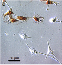
Use histoligical staining by Von Kossa method to measure particle uptake into cells.

In vivo
Use indocyanine green near-infrared imaging to track within body
Transfection Efficiency
Beta-galactosidase GFP-encoding plasmid. (gWiz GFP) Luciferase. Can perform floursecent/lucifer assay, western blot of mouse organs.
