Payne Lab:Research: Difference between revisions
(New page: ='''Imaging Reaction Dynamics in Living Cells'''= Cells are dynamic environments that use carefully regulated mechanisms to maintain function and health. One example of this is the vesicl...) |
No edit summary |
||
| (10 intermediate revisions by 4 users not shown) | |||
| Line 1: | Line 1: | ||
[[Image:PayneBanner.jpg|750px|center]] | |||
<div style="padding: 10px; color: #ffffff; background-color: #000; width: 730px; margin: auto;"> | |||
<center> | |||
[[Payne Lab | <font face="trebuchet ms" style="color:#ffffff"> '''Research''' </font>]] | |||
[[Payne Lab:Contact | <font face="trebuchet ms" style="color:#ffffff"> '''Contact''' </font>]] | |||
[[Payne Lab:Lab Members | <font face="trebuchet ms" style="color:#ffffff"> '''Lab Members''' </font>]] | |||
[[Payne Lab:Reprints | <font face="trebuchet ms" style="color:#ffffff"> '''Publications''' </font>]] | |||
[[Payne Lab:News| <font face="trebuchet ms" style="color:#ffffff"> '''News''' </font>]] | |||
[[Payne Lab:Seminars| <font face="trebuchet ms" style="color:#ffffff"> '''Seminars''' </font>]] | |||
[[Payne Lab:Opportunities| <font face="trebuchet ms" style="color:#ffffff"> '''Opportunities''' </font>]] | |||
</center> | |||
</div><br> | |||
='''Imaging Reaction Dynamics in Living Cells'''= | ='''Imaging Reaction Dynamics in Living Cells'''= | ||
[[Image:cell_rxns_paynelab.gif|right|390 px]] | |||
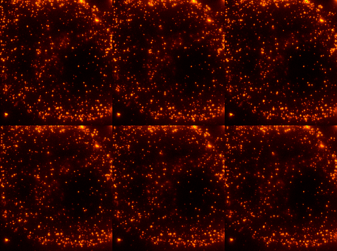
Cells are dynamic environments that use carefully regulated mechanisms to maintain function and health. One example of this is the vesicle-mediated transport of lipids (shown to the right). Each bright spot shows a single vesicle as it transports lipids through the cell. Each step of this process; internalization, transport in the vesicle, and enzymatic degradation of the lipids, is controlled by chemical reactions and mechanical forces within the cell. Understanding these dynamic processes requires a method that will provide both spatial and temporal information-the ability to watch each step as it occurs. To obtain this information we use fluorescence microscopy to directly probe intracellular dynamics. The Payne Lab is interested in two related questions; what are the rates and mechanisms of these intracellular processes and how can we better image each event. | Cells are dynamic environments that use carefully regulated mechanisms to maintain function and health. One example of this is the vesicle-mediated transport of lipids (shown to the right). Each bright spot shows a single vesicle as it transports lipids through the cell. Each step of this process; internalization, transport in the vesicle, and enzymatic degradation of the lipids, is controlled by chemical reactions and mechanical forces within the cell. Understanding these dynamic processes requires a method that will provide both spatial and temporal information-the ability to watch each step as it occurs. To obtain this information we use fluorescence microscopy to directly probe intracellular dynamics. The Payne Lab is interested in two related questions; what are the rates and mechanisms of these intracellular processes and how can we better image each event. | ||
| Line 7: | Line 22: | ||
Direct imaging reveals the subcellular location, concentration, and reaction rate of the molecules of interest. These parameters can be measured to determine a complete intracellular reaction mechanism. Specific systems of interest include post-translational modification of proteins, transcytosis across the blood-brain barrier, and delivery of cargo to the lysosome for degradation. These systems pose a number of biological and physical questions including the mechanism of intracellular transport, kinetics of vesicle fusion, influence of the local environment on a chemical reaction, and the conversion of chemical energy into mechanical motion. | Direct imaging reveals the subcellular location, concentration, and reaction rate of the molecules of interest. These parameters can be measured to determine a complete intracellular reaction mechanism. Specific systems of interest include post-translational modification of proteins, transcytosis across the blood-brain barrier, and delivery of cargo to the lysosome for degradation. These systems pose a number of biological and physical questions including the mechanism of intracellular transport, kinetics of vesicle fusion, influence of the local environment on a chemical reaction, and the conversion of chemical energy into mechanical motion. | ||
==='''New methods for live cell imaging: Fluorescence microscopy and nanomaterial delivery.'''=== | ==='''New methods for live cell imaging: Fluorescence microscopy and nanomaterial delivery.'''=== | ||
The Payne Lab is developing new optical techniques for live cell imaging and new methods for delivering novel fluorescent probes to cells. These methods will be used to probe intracellular reactions on the molecular level and to enable new research directions using quantitative cellular imaging. Optical methods of interest include nanometer-level imaging, spectroscopic single-particle tracking, and multiphoton total internal reflection. The intracellular delivery of novel fluorescent probes will borrow methods developed for gene delivery to introduce fluorescent probes and other nanomaterials into cells in a controlled manner. | The Payne Lab is developing new optical techniques for live cell imaging and new methods for delivering novel fluorescent probes to cells. These methods will be used to probe intracellular reactions on the molecular level and to enable new research directions using quantitative cellular imaging. Optical methods of interest include nanometer-level imaging, spectroscopic single-particle tracking, and multiphoton total internal reflection. The intracellular delivery of novel fluorescent probes will borrow methods developed for gene delivery to introduce fluorescent probes and other nanomaterials into cells in a controlled manner. | ||
Latest revision as of 12:52, 31 July 2009

Imaging Reaction Dynamics in Living Cells

Cells are dynamic environments that use carefully regulated mechanisms to maintain function and health. One example of this is the vesicle-mediated transport of lipids (shown to the right). Each bright spot shows a single vesicle as it transports lipids through the cell. Each step of this process; internalization, transport in the vesicle, and enzymatic degradation of the lipids, is controlled by chemical reactions and mechanical forces within the cell. Understanding these dynamic processes requires a method that will provide both spatial and temporal information-the ability to watch each step as it occurs. To obtain this information we use fluorescence microscopy to directly probe intracellular dynamics. The Payne Lab is interested in two related questions; what are the rates and mechanisms of these intracellular processes and how can we better image each event.
Imaging chemical reactions within a cell.
Direct imaging reveals the subcellular location, concentration, and reaction rate of the molecules of interest. These parameters can be measured to determine a complete intracellular reaction mechanism. Specific systems of interest include post-translational modification of proteins, transcytosis across the blood-brain barrier, and delivery of cargo to the lysosome for degradation. These systems pose a number of biological and physical questions including the mechanism of intracellular transport, kinetics of vesicle fusion, influence of the local environment on a chemical reaction, and the conversion of chemical energy into mechanical motion.
New methods for live cell imaging: Fluorescence microscopy and nanomaterial delivery.
The Payne Lab is developing new optical techniques for live cell imaging and new methods for delivering novel fluorescent probes to cells. These methods will be used to probe intracellular reactions on the molecular level and to enable new research directions using quantitative cellular imaging. Optical methods of interest include nanometer-level imaging, spectroscopic single-particle tracking, and multiphoton total internal reflection. The intracellular delivery of novel fluorescent probes will borrow methods developed for gene delivery to introduce fluorescent probes and other nanomaterials into cells in a controlled manner.