Robert W Arnold Week 6: Difference between revisions
From OpenWetWare
Jump to navigationJump to search
| (14 intermediate revisions by the same user not shown) | |||
| Line 1: | Line 1: | ||
==Electronic Lab Notebook== | ==Electronic Lab Notebook 6== | ||
[[User:Robert W Arnold | Robert W Arnold]] | |||
[[BIOL368/F11:Week 6 | Week 6 Assignment]] | |||
===DNA Glycosylase Exercise=== | ===DNA Glycosylase Exercise=== | ||
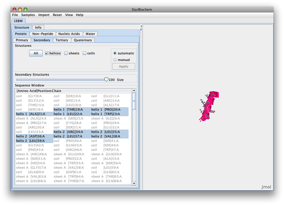
*Downloaded StarBioChem from MIT website. | *Downloaded StarBioChem from MIT website. | ||
*Played around with structure by switching angles and views. | *Played around with structure by switching angles and views. | ||
*Made atoms 100% to | *Made atoms 100% to show accurate space-filled model. | ||
*Began to answer exercise questions. | *Began to answer exercise questions. | ||
* | *Finished up first packet and began to work on Cn3D problems. | ||
===Questions DNA Glycosylase=== | |||
===Questions=== | |||
#Yes, I can identify the DNA and the hOGG1 protein. The DNA is the smaller, double helical structure attached to the space-filled hOGG1 protein. | #Yes, I can identify the DNA and the hOGG1 protein. The DNA is the smaller, double helical structure attached to the space-filled hOGG1 protein. | ||
#One of the amino acid sequences containing a sulfur atom is [CYS]241:A SG #2406. This means it is amino acid: cysteine; position; 241. | #One of the amino acid sequences containing a sulfur atom is [CYS]241:A SG #2406. This means it is amino acid: cysteine; position; 241. | ||
| Line 31: | Line 31: | ||
#Yes, helices are found in hOGG1, they are represented by a purplish pink color. Sheets are also found in hOGG1 and are represented by yellow. Coils are also found in a blueish color in the structure. | #Yes, helices are found in hOGG1, they are represented by a purplish pink color. Sheets are also found in hOGG1 and are represented by yellow. Coils are also found in a blueish color in the structure. | ||
#Amino acids 105-117 all form into a helix structure. Here is the [[Media:Bobby.png | helix]] they form. | #Amino acids 105-117 all form into a helix structure. Here is the [[Media:Bobby.png | helix]] they form. | ||
#The negatively charged amino acids are on the outsides of the protein, exposed. This protein is hydrophilic. | #The negatively charged amino acids are on the outsides of the protein, exposed. This protein is hydrophilic. The helix can be seen [[Media:Picture 7.png | here.]] | ||
#Program continued to freeze when trying to enlarge DNA for this question. Here is the frozen [[Media:Picture 5.png | image.]] | #Program continued to freeze when trying to enlarge DNA for this question. Here is the frozen [[Media:Picture 5.png | image.]] | ||
#Helix 16 would be more likely to recognize damaged guanine bases because at position 315 | #Helix 16 would be more likely to recognize damaged guanine bases because it is directly next to the DNA while Helix 1 is far away at the other side. Helix 16 is also near the glutamine at position 315 which is extremely close to the oxidized gaunine. The two helices can be seen [[Media:Picture 8.png | here.]] Helix 1 and it's side chains can be seen [[Media:Picture 9.png | here.]] and Helix 16 along with it's side chains can be seen [[Media:Picture 10.png | here.]] | ||
===Questions Cn3D=== | |||
#The model of DNA glyosylase in Cn3D is of quaternary structure because there are two polypeptides by determining the ends of the model. | |||
#There are three domains in DNA glycosylase which can be determined by viewed by selecting each one individually; A, C, and D. The Show/Hide structure function is extremely useful for determining this. | |||
#You can view some of the same things, just not nearly as in depth it seems. Cn3D allowed for the structure to be seen as a whole, in parts, but did not allow pieces of the structure to be individually manipulated like we were able to size up certain amino acid chains in StarBioChem. | |||
#Here is a screenshot of the [[Media:Picture 112.png | structure]] in Cn3D. | |||
#Without question, I prefer StarBioChem. The interface was extremely easy to learn and manipulate. I felt comfortable with the program after 10 minutes. For the HIV structure project, I plan to use StarBioChem mostly, but I will also try and incorporate some of Cn3D to diversify a bit and see different representations of comparable information. | |||
==Links== | |||
[[BIOL368/F11 | Biol 368 Homepage]] | |||
{{Robert W Arnold}} | |||
Latest revision as of 15:58, 5 October 2011
Electronic Lab Notebook 6
DNA Glycosylase Exercise
- Downloaded StarBioChem from MIT website.
- Played around with structure by switching angles and views.
- Made atoms 100% to show accurate space-filled model.
- Began to answer exercise questions.
- Finished up first packet and began to work on Cn3D problems.
Questions DNA Glycosylase
- Yes, I can identify the DNA and the hOGG1 protein. The DNA is the smaller, double helical structure attached to the space-filled hOGG1 protein.
- One of the amino acid sequences containing a sulfur atom is [CYS]241:A SG #2406. This means it is amino acid: cysteine; position; 241.
- The sulfur is on the side chain because it remained when backbone was unchecked, but disappeared when side chain was unchecked.
- The 13 amino acids from 105 to 117 are as follows:
- 105 - Threonine
- 106 - Leucine
- 107 - Alanine
- 108 - Glutamine
- 109 - Leucine
- 110 - Tyrosine
- 111 - Histidine
- 112 - Histidine
- 113 - Tryptophan
- 114 - Glycine
- 115 - Serine
- 116 - Valine
- 117 - Aspartic Acid
- Yes, helices are found in hOGG1, they are represented by a purplish pink color. Sheets are also found in hOGG1 and are represented by yellow. Coils are also found in a blueish color in the structure.
- Amino acids 105-117 all form into a helix structure. Here is the helix they form.
- The negatively charged amino acids are on the outsides of the protein, exposed. This protein is hydrophilic. The helix can be seen here.
- Program continued to freeze when trying to enlarge DNA for this question. Here is the frozen image.
- Helix 16 would be more likely to recognize damaged guanine bases because it is directly next to the DNA while Helix 1 is far away at the other side. Helix 16 is also near the glutamine at position 315 which is extremely close to the oxidized gaunine. The two helices can be seen here. Helix 1 and it's side chains can be seen here. and Helix 16 along with it's side chains can be seen here.
Questions Cn3D
- The model of DNA glyosylase in Cn3D is of quaternary structure because there are two polypeptides by determining the ends of the model.
- There are three domains in DNA glycosylase which can be determined by viewed by selecting each one individually; A, C, and D. The Show/Hide structure function is extremely useful for determining this.
- You can view some of the same things, just not nearly as in depth it seems. Cn3D allowed for the structure to be seen as a whole, in parts, but did not allow pieces of the structure to be individually manipulated like we were able to size up certain amino acid chains in StarBioChem.
- Here is a screenshot of the structure in Cn3D.
- Without question, I prefer StarBioChem. The interface was extremely easy to learn and manipulate. I felt comfortable with the program after 10 minutes. For the HIV structure project, I plan to use StarBioChem mostly, but I will also try and incorporate some of Cn3D to diversify a bit and see different representations of comparable information.
Links
- Robert W Arnold Week 2
- Robert W Arnold Week 3
- Robert W Arnold Week 4
- Robert W Arnold Week 5
- Robert W Arnold Week 6
- Robert W Arnold Week 7
- Robert W Arnold Week 8
- Robert W Arnold Week 9
- Robert W Arnold Week 10
- Robert W Arnold Week 11
- Robert W Arnold Week 12
- Robert W Arnold Week 14
- Week 2 Assignment
- Week 3 Assignment
- Week 4 Assignment
- Week 5 Assignment
- Week 6 Assignment
- Week 7 Assignment
- Week 8 Assignment
- Week 9 Assignment
- Week 10 Assignment
- Week 11 Assignment
- Week 12 Assignment
- Week 14 Assignment
- Class Journal Week 1
- Class Journal Week 2
- Class Journal Week 3
- Class Journal Week 4
- Class Journal Week 5
- Class Journal Week 6
- Class Journal Week 7
- Class Journal Week 8
- Class Journal Week 9
- Class Journal Week 10
- Class Journal Week 11
- Class Journal Week 12
- Class Journal Week 14