Samantha M. Hurndon Week 6: Difference between revisions
From OpenWetWare
Jump to navigationJump to search
(New page: ==DNA Glycosylase== ==Intro== *We will explore the relationship between a protein’s structure and its function in a human DNA glycosylase (hOGG1) ==Methods== #I first got started by op...) |
(No difference)
|
Revision as of 15:37, 5 October 2011
DNA Glycosylase
Intro
- We will explore the relationship between a protein’s structure and its function in a human DNA glycosylase (hOGG1)
Methods
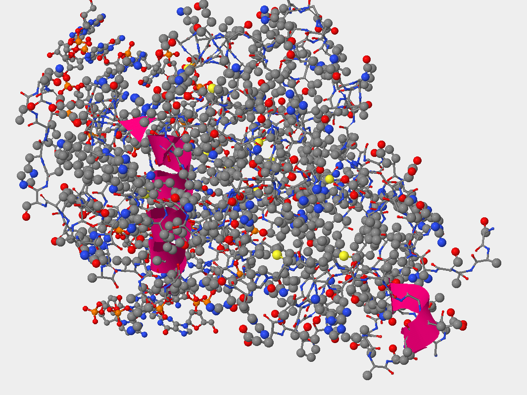
- I first got started by opening the program StarBiochem and opened the Sample “DNA glycosylase hOGG! w/ DNA – H. sapiens (1EBM)”
- The structure was in a ball-and-stick model, which Is the defult in biochem. This allows us to see how atoms of the structure are bonded together. Although that is a nice thing to see we want to see the space each atom fills.
- We did this by looking at the space-filled model, here size is represented of the physical space each atom occupies.
- A series of questions was completed below:
Exercise
- Can you identify the DNA and the hOGG1 protein in the structure. Where is the DNA segment?
- In the picture below you can see that the DNA occupies less space than the protein atoms. Also, what is seen is the double helix that indicates DNA.
- hOGG1 contains multiple sulfur atoms.
- Identify the name and sequence number of one of the amino acids in the structure that contains a sulfur atom.
- [CYS]241:ASG#2406
- Identify the name and sequence number of one of the amino acids in the structure that contains a sulfur atom.
- The sulfur atom is located in the side chain region of the amino acid, as seen below.
- Primary structure of the hOGG1 protein. Amino acids 105-117 complete amino acid name
- 105: Threonine
- 106: Leucine
- 107: Alanine
- 108:Glutamine
- 109: Leucine
- 110: Tyrosine
- 111: Histidine
- 112: Histidine
- 113: Tryptophan
- 114: Glycine
- 115: Serine
- 116: Valine
- 117: Aspartic acid
- Protein chain has amino acids that form local structures called secondary structures.
- Are helices, sheets or coils present in hOGG1? Describe the color that represents each secondary structure you observe.
- I observed helices, sheets and coils were all present (photo shown below)
- Which secondary structure does amino acids 105-117 fold into?
- They fold into a helice structure (shown below)
- Are helices, sheets or coils present in hOGG1? Describe the color that represents each secondary structure you observe.
- Looking at DNA glycosylase’s structure in relation to tertiary structure.
- Negatively charged amino acids are located on the outside (exposed) of this protein. Because it is known that negatively charged amino acids are hydrophilic, and it is observed that the negatively charged amino acids (pink color) are exposed to the outside environment, this indicates that these negatively charged amino acids wants to interact with the environment. (look at photo below)
- hOGG1’s interaction with DNA


