Sean Lauber:Differential cell staining: Difference between revisions
| (12 intermediate revisions by the same user not shown) | |||
| Line 34: | Line 34: | ||
'''Macrophage/Monocyte:''' | '''Macrophage/Monocyte:''' | ||
- Large cells; monocytes are smaller | - Large cells; monocytes are smaller | ||
- Dark Staining nucleus | - Dark Staining nucleus | ||
- Large cytoplasm that staisn clear or light purple; monocytes have a smaller cytoplasm | - Large cytoplasm that staisn clear or light purple; monocytes have a smaller cytoplasm | ||
- Frizzled outer membrane (macrophage processes) when activated; Monocytes have a smoother outer membrane | - Frizzled outer membrane (macrophage processes) when activated; Monocytes have a smoother outer membrane | ||
- Can have lots of vessicles inside | - Can have lots of vessicles inside | ||
- Sometimes multinucleated (when close to dividing or when recently engulfed a cell) | - Sometimes multinucleated (when close to dividing or when recently engulfed a cell) | ||
'''Lymphocyte (look very similar to monocytes so be careful):''' | '''Lymphocyte (look very similar to monocytes so be careful):''' | ||
- Small cell | - Small cell | ||
- Dark staining nucleus | - Dark staining nucleus | ||
- Have very little cytoplasm (a tiny slit) (monocytes tend to have more) that stains clear or light purple | - Have very little cytoplasm (a tiny slit) (monocytes tend to have more) that stains clear or light purple | ||
- Sometimes all you see is a dark staining nucleus, if you look closely you'll see the membrane containing very little cytoplasm | - Sometimes all you see is a dark staining nucleus, if you look closely you'll see the membrane containing very little cytoplasm | ||
- Can be frizzled | - Can be frizzled | ||
'''Neutrophil''' | '''Neutrophil''' | ||
- Small cell | - Small cell | ||
- Dark staining, lobulated nucleus | - Dark staining, lobulated nucleus | ||
- Has little cytoplasm that stains clear or light purple | - Has little cytoplasm that stains clear or light purple | ||
'''Eosinophil''' | '''Eosinophil''' | ||
- Small cell | - Small cell | ||
- Dark staining, lobulated nucleus | - Dark staining, lobulated nucleus | ||
- Has little cytoplasm with granules that stain pink | - Has little cytoplasm with granules that stain pink | ||
[http://openwetware.org/images/e/e3/Differential1.jpg Example 1] | |||
[http://openwetware.org/images/4/48/Differential2.jpg Example 2] | |||
[http://openwetware.org/images/4/4d/Differential3.jpg Example 3] | |||
[http://openwetware.org/images/d/d9/Differential4.jpg Dominik's notes] | |||
Latest revision as of 12:51, 30 October 2012
To differentially stain the cytocentrifuge smears (from BAL), the Hema3 reagent is used (Fisher, 23-123-869).
Protocol for staining (each dip is done for 1 second):
1. Dip one slide into the fixative (clearish blue) 5times.
2. Drain excess fixative.
3. Dip slide into the Xanthene solution (red) 4 times.
4. Blot onto paper towel.
5. Dip slide into the Thiazine solution (purple) 3 times.
6. Drain excess.
7. Rinse the slide immediately with distilled water.
8. Allow the slide to air dry completely overnight.
9. Mount with Permount or something similar and coverslip.
If a deeper stain is desired, add another dipping step. Remove a step is you want less staining.
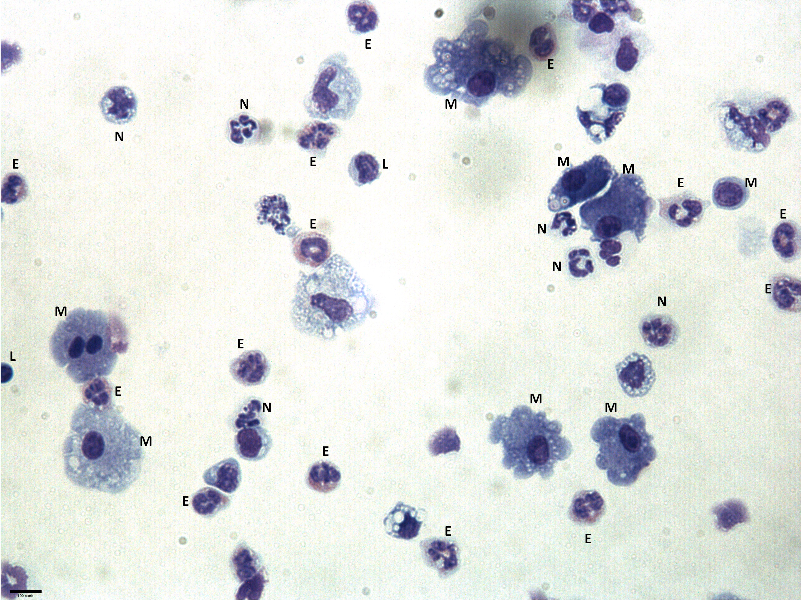
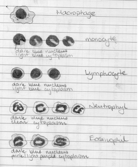
Differential counting
Count up to 500 cells for each cytocentrifuge smear and classify each cell as either a macrophage/monocyte, lymphocyte, neutrophil or eosinophil. Then calculate the % for each from the total. Then you can apply the % to the total cell count to determine the total number of each of these cell types. This will let you know if certain cell types are increased upon treatment.
Macrophage/Monocyte:
- Large cells; monocytes are smaller
- Dark Staining nucleus
- Large cytoplasm that staisn clear or light purple; monocytes have a smaller cytoplasm
- Frizzled outer membrane (macrophage processes) when activated; Monocytes have a smoother outer membrane
- Can have lots of vessicles inside
- Sometimes multinucleated (when close to dividing or when recently engulfed a cell)
Lymphocyte (look very similar to monocytes so be careful):
- Small cell
- Dark staining nucleus
- Have very little cytoplasm (a tiny slit) (monocytes tend to have more) that stains clear or light purple
- Sometimes all you see is a dark staining nucleus, if you look closely you'll see the membrane containing very little cytoplasm
- Can be frizzled
Neutrophil
- Small cell
- Dark staining, lobulated nucleus
- Has little cytoplasm that stains clear or light purple
Eosinophil
- Small cell
- Dark staining, lobulated nucleus
- Has little cytoplasm with granules that stain pink