User:Brendan A Norwood-Pearson/Notebook/Biology 210 at AU: Difference between revisions
Sarah Knight (talk | contribs) No edit summary |
No edit summary |
||
| Line 1: | Line 1: | ||
"16s Sequence from Bacteria Lab- March 3 2015" | |||
'''Purpose'''- The purpose of this experiment was for the lab to be able to take a closer look at the PCR products that we ran a while ago. The Gene that we amplified was the 16s gene from our bacteria that we grew from our transect samples. Looking at the products of the sanger sequencing and blasting them will ultimately enable the lab to identify the species of Bacteria and report on it. | |||
'''Materials and Methods'''- The lab used raw sequences of our 16s gene per amplifications and put it into the BLAST system. Blast stands for Basic Local Alignment Search Tool. What it does is takes a sequence and matches it to an organism's genetic make-up as close as possible. By simply entering the data that was collected from sanger sequencing into the BLAST data base, the lab was able to identify our bacteria species. | |||
Results- Listed Below is the raw sequence of our sample. | |||
MB 35: '''Chryseobacterium''' NNNNNNNNNNNNNGCTNNGCAGCCGAGCGGTAGAGATTCTTCGGAATCTTGAGAGCGGCGTACGGGTGCGGAACACGTGT GCAACCTGCCTTTATCAGGGGGATAGCCTTTCGAAAGGAAGATTAATACCCCATAATATATTAAGTGGCATCACTTGATA TTGAAAACTCCGGTGGATAAAGATGGGCACGCGCAAGATTAGATAGTTGGTAGGGTAACGGCCTACCAAGTCAGTGATCT TTAGGGGGCCTGAGAGGGTGATCCCCCACACTGGTACTGAGACACGGACCAGACTCCTACGGGAGGCAGCAGTGAGGAAT ATTGGACAATGGGTGAGAGCCTGATCCAGCCATCCCGCGTGAAGGATGACGGCCCTATGGGTTGTAAACTTCTTTTGTAT AGGGATAAACCTTTCCACGTGTGGAAAGCTGAAGGTACTATACGAATAAGCACCGGCTAACTCCGTGCCAGCAGCCGCGG TAATACGGAGGGTGCAAGCGTTATCCGGATTTATTGGGTTTAAAGGGTCCGTAGGCGGATCTGTAAGTCAGTGGTGAAAT CTCACAGCTTAACTGTGAAACTGCCATTGATACTGCAGGTCTTGAGTAAGGTAGAAGTAGCTGGAATAAGTANTGTAGCG GTGAAATGCATANATATTACNTANNNNACCAATTGCGAAGGCAGGTTACTATGTCTTAACTGACNCTGATGGACNAAAGC GTGGGGAGCGAACAGGATTNNATACCCTGGTAGTCCACNCCGTAAACGATGCTAACTCGTTTTTGGGNCTTCGGATTCNN ANACTAANCGAAAGTGATAAGTTAGCCNACCTGGGAGTACNGTTNNNNAGAATGAAACTNNNANNANNNACGGGGGCCNN NNNNACCGGNNNNTATNTGGTTTNAGTGCNATGATNCGCGAGNANCNNTACCACGCNTNAANTGGGGAAGTGAGGNNGGT TNGNNNGNNNNCGTNNNTNCNNCAANTTTCAANNNNNTCCNGGGTGNNGGNGGGNNGTNNNNNANNGNTANGTNTAGGTN CNNNGACNAGCNCNNCCCCCNNNNNCGGGNNNGGGCGGNTNNNTTNCNGGANNNCNNNNNGNGNNNCCCTTGNNNNN | |||
MB 36: '''Variovarax''' NNNNNNNNNNNNNNNNNTNNNNTGCAGTCGTANGGNGGTCAGCGCGTANCAATCCTGGCGGCGAGTGGCGAACGGGTGAG TAATACATCGGAACGTGCCCAATCGTGGGGGATAACGCAGCGAAAGCTGTGCTAATACCGCATACGATCTACGGATGAAA GCAGGGGATCGCAAGACCTTGCGCGAATGGAGCGGCCGATGGCAGATTAGGTAGTTGGTGAGGTAAAGGCTCACCAAGCC TTCGATCTGTAGCTGGTCTGAGAGGACGACCAGCCACACTGGGACTGAGACACGGCCCAGACTCCTACGGGAGGCAGCAG TGGGGAATTTTGGACAATGGGCGAAAGCCTGATCCAGCCATGCCGCGTGCAGGATGAAGGCCTTCGGGTTGTAAACTGCT TTTGTACGGAACGAAACGGCCTTTTCTAATAAAGAGGGCTAATGACGGTACCGTAAGAATAAGCACCGGCTAACTACGTG CCAGCAGCCGCGGTAATACGTAGGGTGCAAGCGTTAATCGGAATTACTGGGCGTAAAGCGTGCGCAGGCGGTTATGTAAG ACAGTTGTGAAATCCCCGGGCTCAACCTGGGAACTGCATCTGTGACTGCATAGCTAGAGTACGGTAGAGGGGGATGGGAA TTCCGCGTGTAGCAGTGAAATGCGTAGATATGCGGAGGAACACCGATGGCGAAGGCAATCCCCTGGACCTGTACTGACGC TCATGCACGAAANNNGTGGGGAGCAAACAGGATTANATACCCTGGTAGTCCACGCCCTAAACGATGTCNACTGGTTGTTG GGTCTTCACTGACTCANTAACNAANCTAACNCGTGAAGTTGACCGCCTGGGGANTACGGCCGCAAGGTTGNAAACTCANN GAATTGANNGGGNNCCNCACAANCNGTGGATGATGTGGTTTATTTNNTNNCAACGCNAAAACCTTACCCACCTTTNGACA TNTACGGAATTCGCCNGANATGGNTTANTGCTCGANAGNANAANCNNTACANCNNGTGCTGCATGGCTGTNNTCNNCTCG TGTNNNTGNNANNNTGGNTNANNCCCNCAACNNNCGCAACCCNNNNTCATTNNNTGGCTACNTTCGNNTNGGCNNTCTNN GGNNACTGCCNGNTGANNNAC | |||
The species identified as Variovarax and Chryseobacterium. | |||
[[Image:IMG 1236.jpg]] | |||
Shown above is a picture of the gel that was originally run. Indicated by the red circle is the two gene samples that were sent in for sanger sequencing. | |||
'''Results''' - | |||
The Variovarax genus is a family of bacteria that can be found in soil. From this, the lab concluded that this type of bacteria came from our ground litter sample that we extracted from our transect. Bacteria in this genus have been long known to break down boron, a toxic element that can be harmful to certain aspects of ecosystems similar to that of our transect. | |||
The Chryseobacterium bacteria is a member of a family of bacteria that is closely linked to meningitis. This bacteria has no mention of being able to directly benefit the type of ecosystem that we collected our data from, but it is actually very cool to see the diversity of such bacteria in a small place like a transect at AU. | |||
'''Work Cited''' | |||
Jacobs, R., KC Bloch, and R. Nadarajah. "Chryseobacterium Meningosepticum: An Emerging Pathogen among Immunocompromised Adults. Report of 6 Cases and Literature Review." NCBI (1997): n. pag. Pubmed.com. Web. 2 Mar. 2015. | |||
H. Miwa - I. Ahmed - J. Yoon - A. Yokota - T. Fujiwara - International Journal Of Systematic And Evolutionary Microbiology - 2008 | |||
'''Zebra Fish Experiment- Feburary 25 2015''' | '''Zebra Fish Experiment- Feburary 25 2015''' | ||
Revision as of 22:53, 1 March 2015
"16s Sequence from Bacteria Lab- March 3 2015"
Purpose- The purpose of this experiment was for the lab to be able to take a closer look at the PCR products that we ran a while ago. The Gene that we amplified was the 16s gene from our bacteria that we grew from our transect samples. Looking at the products of the sanger sequencing and blasting them will ultimately enable the lab to identify the species of Bacteria and report on it.
Materials and Methods- The lab used raw sequences of our 16s gene per amplifications and put it into the BLAST system. Blast stands for Basic Local Alignment Search Tool. What it does is takes a sequence and matches it to an organism's genetic make-up as close as possible. By simply entering the data that was collected from sanger sequencing into the BLAST data base, the lab was able to identify our bacteria species.
Results- Listed Below is the raw sequence of our sample.
MB 35: Chryseobacterium NNNNNNNNNNNNNGCTNNGCAGCCGAGCGGTAGAGATTCTTCGGAATCTTGAGAGCGGCGTACGGGTGCGGAACACGTGT GCAACCTGCCTTTATCAGGGGGATAGCCTTTCGAAAGGAAGATTAATACCCCATAATATATTAAGTGGCATCACTTGATA TTGAAAACTCCGGTGGATAAAGATGGGCACGCGCAAGATTAGATAGTTGGTAGGGTAACGGCCTACCAAGTCAGTGATCT TTAGGGGGCCTGAGAGGGTGATCCCCCACACTGGTACTGAGACACGGACCAGACTCCTACGGGAGGCAGCAGTGAGGAAT ATTGGACAATGGGTGAGAGCCTGATCCAGCCATCCCGCGTGAAGGATGACGGCCCTATGGGTTGTAAACTTCTTTTGTAT AGGGATAAACCTTTCCACGTGTGGAAAGCTGAAGGTACTATACGAATAAGCACCGGCTAACTCCGTGCCAGCAGCCGCGG TAATACGGAGGGTGCAAGCGTTATCCGGATTTATTGGGTTTAAAGGGTCCGTAGGCGGATCTGTAAGTCAGTGGTGAAAT CTCACAGCTTAACTGTGAAACTGCCATTGATACTGCAGGTCTTGAGTAAGGTAGAAGTAGCTGGAATAAGTANTGTAGCG GTGAAATGCATANATATTACNTANNNNACCAATTGCGAAGGCAGGTTACTATGTCTTAACTGACNCTGATGGACNAAAGC GTGGGGAGCGAACAGGATTNNATACCCTGGTAGTCCACNCCGTAAACGATGCTAACTCGTTTTTGGGNCTTCGGATTCNN ANACTAANCGAAAGTGATAAGTTAGCCNACCTGGGAGTACNGTTNNNNAGAATGAAACTNNNANNANNNACGGGGGCCNN NNNNACCGGNNNNTATNTGGTTTNAGTGCNATGATNCGCGAGNANCNNTACCACGCNTNAANTGGGGAAGTGAGGNNGGT TNGNNNGNNNNCGTNNNTNCNNCAANTTTCAANNNNNTCCNGGGTGNNGGNGGGNNGTNNNNNANNGNTANGTNTAGGTN CNNNGACNAGCNCNNCCCCCNNNNNCGGGNNNGGGCGGNTNNNTTNCNGGANNNCNNNNNGNGNNNCCCTTGNNNNN
MB 36: Variovarax NNNNNNNNNNNNNNNNNTNNNNTGCAGTCGTANGGNGGTCAGCGCGTANCAATCCTGGCGGCGAGTGGCGAACGGGTGAG TAATACATCGGAACGTGCCCAATCGTGGGGGATAACGCAGCGAAAGCTGTGCTAATACCGCATACGATCTACGGATGAAA GCAGGGGATCGCAAGACCTTGCGCGAATGGAGCGGCCGATGGCAGATTAGGTAGTTGGTGAGGTAAAGGCTCACCAAGCC TTCGATCTGTAGCTGGTCTGAGAGGACGACCAGCCACACTGGGACTGAGACACGGCCCAGACTCCTACGGGAGGCAGCAG TGGGGAATTTTGGACAATGGGCGAAAGCCTGATCCAGCCATGCCGCGTGCAGGATGAAGGCCTTCGGGTTGTAAACTGCT TTTGTACGGAACGAAACGGCCTTTTCTAATAAAGAGGGCTAATGACGGTACCGTAAGAATAAGCACCGGCTAACTACGTG CCAGCAGCCGCGGTAATACGTAGGGTGCAAGCGTTAATCGGAATTACTGGGCGTAAAGCGTGCGCAGGCGGTTATGTAAG ACAGTTGTGAAATCCCCGGGCTCAACCTGGGAACTGCATCTGTGACTGCATAGCTAGAGTACGGTAGAGGGGGATGGGAA TTCCGCGTGTAGCAGTGAAATGCGTAGATATGCGGAGGAACACCGATGGCGAAGGCAATCCCCTGGACCTGTACTGACGC TCATGCACGAAANNNGTGGGGAGCAAACAGGATTANATACCCTGGTAGTCCACGCCCTAAACGATGTCNACTGGTTGTTG GGTCTTCACTGACTCANTAACNAANCTAACNCGTGAAGTTGACCGCCTGGGGANTACGGCCGCAAGGTTGNAAACTCANN GAATTGANNGGGNNCCNCACAANCNGTGGATGATGTGGTTTATTTNNTNNCAACGCNAAAACCTTACCCACCTTTNGACA TNTACGGAATTCGCCNGANATGGNTTANTGCTCGANAGNANAANCNNTACANCNNGTGCTGCATGGCTGTNNTCNNCTCG TGTNNNTGNNANNNTGGNTNANNCCCNCAACNNNCGCAACCCNNNNTCATTNNNTGGCTACNTTCGNNTNGGCNNTCTNN GGNNACTGCCNGNTGANNNAC
The species identified as Variovarax and Chryseobacterium.
Shown above is a picture of the gel that was originally run. Indicated by the red circle is the two gene samples that were sent in for sanger sequencing.
Results -
The Variovarax genus is a family of bacteria that can be found in soil. From this, the lab concluded that this type of bacteria came from our ground litter sample that we extracted from our transect. Bacteria in this genus have been long known to break down boron, a toxic element that can be harmful to certain aspects of ecosystems similar to that of our transect.
The Chryseobacterium bacteria is a member of a family of bacteria that is closely linked to meningitis. This bacteria has no mention of being able to directly benefit the type of ecosystem that we collected our data from, but it is actually very cool to see the diversity of such bacteria in a small place like a transect at AU.
Work Cited
Jacobs, R., KC Bloch, and R. Nadarajah. "Chryseobacterium Meningosepticum: An Emerging Pathogen among Immunocompromised Adults. Report of 6 Cases and Literature Review." NCBI (1997): n. pag. Pubmed.com. Web. 2 Mar. 2015.
H. Miwa - I. Ahmed - J. Yoon - A. Yokota - T. Fujiwara - International Journal Of Systematic And Evolutionary Microbiology - 2008
Zebra Fish Experiment- Feburary 25 2015
For the zebra fish experiment my partner is Roshni. We both decided to use the Estrogen as our independent variable. We hypothesize that the presence of estrogen will cause the zebra fish to grow at a faster rate. More specifically, their body mass will be disproportional to their organ mass in comparison to the control zebra fish. So far we have growing zebra fish in our petri dishes. One control and one test. The test is filled with 25mL of estrogen and the control is filled with 25mL of control water. We will record their size and average them out along the course of a couple of weeks. This is cool.
BNP
2.24.15
Good Invertebrate entry. Missing the vertebrate section with description of vertebrates present in transect (or potentially present) and a food web.
SK
" Invertebrates-Feburary 18 2015"
Purpose The purpose of this lab was to look at the different invertebrates collected from our Berlese Funnel and to look at other invertebrates in order to experience the diversity of the invertebrate family first hand. The lab recorded the phylum, size, quantity of the invertebrates within the petri dish, and a description for each of the different invertebrates that we observed.
Methods and Materials The invertebrates were collected out of the ethanol that they fell into in our Berlese Funnel and put into a clear petri dish for viewing via microscope. The results were nothing short of overwhelming as many different type of invertebrates were discovered. A centipede, a small sole mite, a spring tail, and a large soil mite were all found in our transect.
Results
Above is the data that was collected.A centipede, a small sole mite, a spring tail, and a large soil mite were all found in our transect. They ranged from .2 to 1mm in size. The lab recorded the phylum, size, quantity of the invertebrates within the petri dish, and a description for each of the different invertebrates that we observed.
Above is the centipede that was observed by the lab. It was identified by the number of legs and unique appearance. This was a very exciting find for the lab.
Discussion
Additionally, an acoelomat, a planaria, and a nematode were observed. The acoleomat was observed as moving with its whole body. It seemed as if the worm used a lot of energy to go a short distance. The planaria was observed to move more like a snake, very slow and decisively. The nematode was seen to slither.
The invertebrates observed have been shown to participate in many different aspects of ecology. For example the centipede is crucial to the ecosystem by being a predator pushing natural selection, and soil mites have been shown to break down dead leaves and release the nutrients back into the soil. In all, the invertebrates observed are shown to carry their weight in the ecology system.
Conclusion and Future Directions In conclusion the invertebrates that the lab observed showed to effect both biotic and abiotic aspects of our transects. Next week we will begin our trials with zebra fish!
BNP
2.19.15 Good lab book entry. Focus should be on the plant part of the lab as the PCR part will be more relevant when get sequence data back. SK
"PCR for 16s along with Plantae and Fungi-Febuary 11 2015 "
"Purpose" The purpose of this experiment was to put the PCR product that accumulated in last week's lab and run it through a gel. We also returned back to our transect spot, where we collected samples for our Hay Infusion Culture, and picked leaves off of trees and bushes. We then used our knowledge obtained in class and in the lab manual to jot down various characteristics about the leaves like vascularization and mechanism of reproduction, just to name a few. Finally, we needed samples from our transect in order to set up a Berlese Funnel to Collect Invertebrates.
"Materials and Methods" We used the PCR product from last lab, combined 5 micro liters of our sample with 3 micro liters of dye, and pipetted it into a gel electrophoresis well. After running the gel, we recorded the data. The lab visited our transect sight and got samples of ground material and 5 samples of leaves. We observed the leaves and put in ground material in an inverted funnel with a filter known as a Berlese Funnel.
Data and Observations
Above is a picture of the PCR gel that we ran to amplify the 16s Gene. As you can see, in the first well (from left to right) we added a ladder to measure the base pairs of the gene, and each PCR product was put in a well.
 Above are the data for the five leaves that we collected. Characteristics include size, vascularization, location, specialized structures, and Mechanisms of Reproduction. We found our leaves on trees and pushes, some of them were dead and had fallen off. As it turns out, just like humans have vascularization, leaves have vascularization too. We found xylem and phloem under the belly of our leaves which indicated that they all had vascularization. We also saw cuticles, a layer of waxy substance, on the outer edge of our leaves too.
---Leaf 1 came from a low hanging plant in a loose cluster of other leaves
---Leaf 2 came from a bush with dense leaf clusters
---Leaf 3 came from the ground
---Leaf 4 came from a tall tree with dense leaf clusters
---Leaf 5 came from a small bush with a loose leaf cluster
No fungi were found in our transect. Fungi sporangia are important because they produce haploid spores or "seeds" that give fungi the ability to grow in other places.
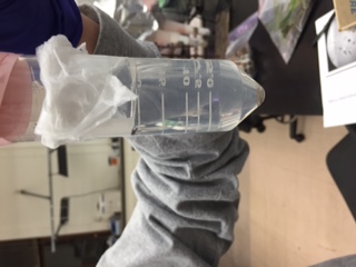
Above is a picture of the Berlese Funnel, I assume that we are going to be using it in order to examine Invertebrates in the near future.
2.10.15 Very good entry. Could include serial dilution data table in results section. Address all red text in manual. Also give some detail about the gram stain protocol or include a link to a comparable protocol. SK
Microbiology and Identifying Bacteria with DNA Sequences- January 29 2015
Purpose To build off of the knowledge obtained from last lab section by observing the different characteristics of bacteria, experience the reality of antibiotic resistance, and to use DNA sequences in order to identify different species of bacteria.
Materials and Methods The lab looked at each of the petri dishes containing the bacteria from the Hay Infusion Culture and recorded evidence of antibiotic resistance. In order to observe the bacteria that we grew last lab section, we extracted samples from each petri dish, made a wet mount, and looked at it through the microscope. We then stained the bacteria we observed in order to determine weather the bacteria were gram negative or positive. Finally, we isolated DNA from the bacteria and amplified the 16S rNA gene via PCR for our next lab.
Data and Observations First off, our Hay Infusion Culture had a lot less water inside of it. More than likely due to evaporation. It smelled a lot worse in comparison to the last time we took samples from it. There was also mold present within it. We observed that the platelets with the tetracycline antibiotic yielded less bacteria colonies than the ones without it. This was expected due to the fact that tetracycline is known to prohibit the bacteria polymerase from continuing transcription. However, we also found that a purple colored colony seemed to be persistent throughout all of the dishes suggesting that it may have a resistance to the tetracycline antibiotic.
 This is a Gram Negative Spirillum that we found within our Hay Infusion Culture. Its pinkish color indicates that it does not have an outer layer made of peptidoglycan (x100 magnification)
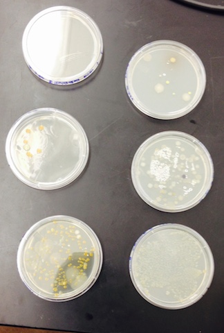
 This is a picture of the bacteria colonies that we collected samples from and observed the antibiotic resistance.

For the PCR reaction we used two samples from the 10x7 Tet colony and two samples from the 10x9 Tet colony.
Conclusions and Future Directions In conclusion, we observed evidence of the Tetracycline antibiotic working as an antibiotic to some colonies, and we also witnessed resistance against it. Through our observations via microscope we were able to identify different characteristics and types of bacteria in our Hayford Infusion Culture and determine weather they were gram negative or positive via staining. When we return to the lab, we will use the amplified 16S gene and put it through a gel. Once the samples from the Gel or sequenced, we will use them to identify bacteria.
BNP
2.4.15 Good notebook entry. Picture is too big. Try saving it as a smaller file size and then uploading it. SK
Hay Infusion Culture Observations and Preparing and Plating Serial Dilutions- January 22 2015
Purpose In order to apply the knowledge learned last week to this week, examination of samples from our previously prepared Hay Infusion Cultures is necessary. The lab has reasons to believe that there are unicellular Eukaryotic organisms within our sample and we will identify them. Plating serial dilutions need to be done in order to prepare for next week's lab.
Materials and Methods A sample of the Hay Infusion Culture was taken and observed through a microscope and many different types of organisms were found within the sample. A Dichotomous key was used in order to identify different organisms and the characteristics that they embody. A wet mount of known organisms were observed first in order to familiarize ourselves with some of the unicellular organisms that we could see in our sample. The results were recorded. For the serial dilutions, 10 uL of the Hay infusion sample was taken and put through a three series 100 ul dilution with sterile broth. From those dilutions, 100 ul of each of the tubes within the dilution series was transferred into nutrient agar plates. A separate 100 ul of original dilution series was put into a nutrient agar plus tetracycline plate. This was done so that bacteria colonies would grow. The details of this dilution are depicted below.

Data and Observations Different organisms were living at different levels of our Hay infusion Culture so the lab took samples from the top, the middle, and the bottom. At the top we found Chlamydomnas (10 micrometers), Clopidiunm (50 micrometers), and Paramecium (90 micrometers). At the bottom of the sample we found Peranama (50 micrometers), Chalmydonas (7 micrometers), and Paramecium Caudatum (230 micrometers). In the middle Paramecium bursaria (90 micrometers) were found. One organism that caught the interest of the lab was the Chlamydomnas and its intense flagella that enabled the organism to be very fast and motile. It was difficult to identify the organism because of its speed. Luckily, we caught it as it was taking a breather.
Conclusions and Future Directions From this lab, we can conclude that the diversity of organisms within our transect is very colorful in regards to types of organisms. If our sample were to grow for another two months, we would probably observe a lot more kids of organisms present. We would need a lot of proteasome!
Observation of Volvocine Line and Observing a Niche at Au- January 15th 2015
Purpose: To examine organism diversity as a product of evolution first hand by looking at the Volvocine Line algae group through the microscope. Then, venture out into the natural habitat of these organisms by taking samples of soil from our assigned 20x20 transect. From our samples, we will differentiate between abiotic and biotic components within our niche and create a Hay Infusion Culture for future projects.
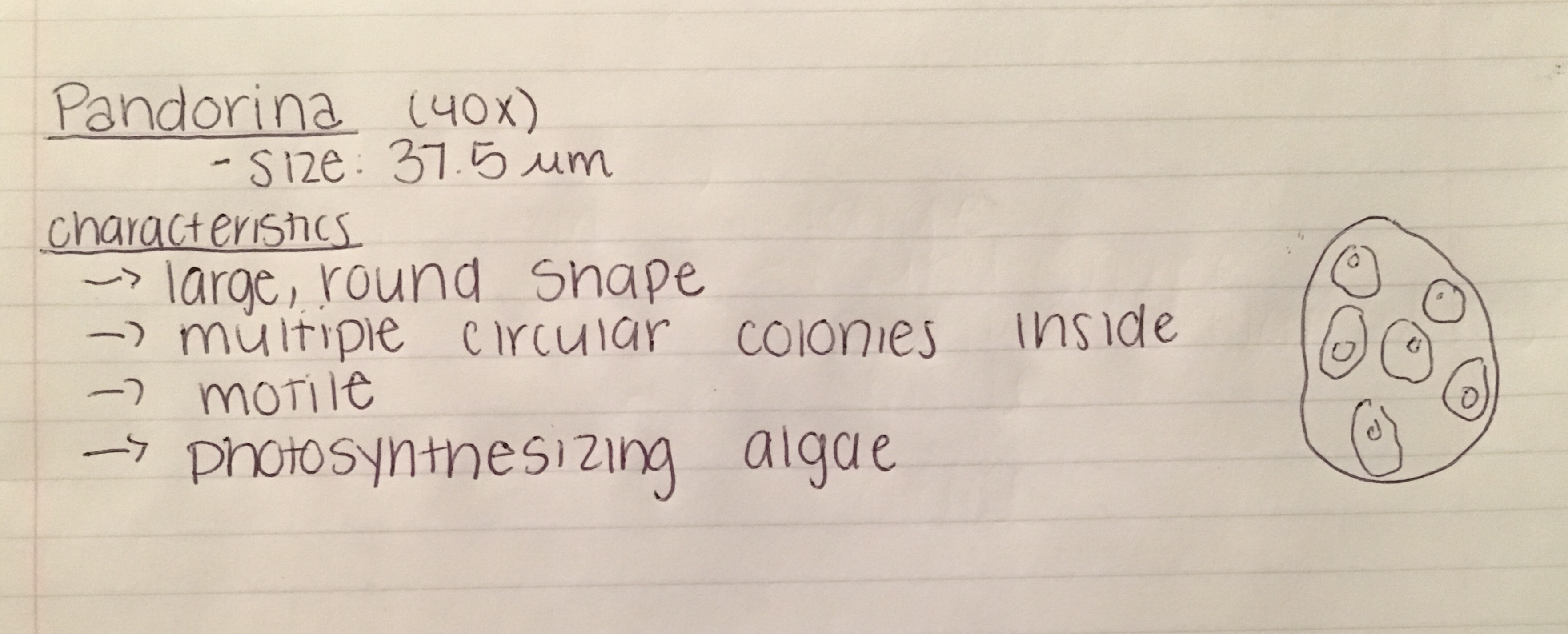
Materials and Methods: As a class, we used Microscopes and hand prepared slides in order to look at the organisms of interest. They were Chlamydomas, Gonium, and Volvox collectively. After recording the number of cells, colony size, specialization, mechanisms of motility, and the status of their gametes we were able to get a sense of the diversity of just one type of organism. With this in mind, the lab journeyed out to our assigned transect and collected aspects of our transects and put it in a tube. This tube was later emptied into a jar along with some milk and water, and saved on a shelf.
This is a sketch of the transect that the sample for the Hay Infusion was taken from. It was a very lush corner of our campus with lots of plants and bushes. Abiotic components consisting of dirt, rocks, snow, a lamp post, and metal signs. Biotic components consisting of trees, ants, bushes, micro organisms, birds.
This is the collected data from the Volvocine Line.
Conclusion and Future Directions: We can conclude that evolution has indeed made our world diverse in the organism realm. In the future, we will look at the Hay infusion Culture and collect samples from it.
BNP


