User:Corey J Salas/Notebook/Biology 210 at AU: Difference between revisions
No edit summary |
|||
| (146 intermediate revisions by 2 users not shown) | |||
| Line 1: | Line 1: | ||
== Bacterial PCR Data == | |||
'''PURPOSE''': The purpose of the PCR lab was to identify the type of bacteria present in the assigned transect by isolation and amplification of a single gene 16S. | |||
<br> | |||
<br>'''MATERIALS AND METHODS''': | |||
PCR for 16S sequencing was set up using one of the four different bacterial colonies in the hay infusion culture. A sample of the bacteria in the colony was transferred into a 100μL sterile tube, incubated at 100°C for 10 minutes, centrifuged for 5 minutes at 13,400rpm, mixed with a primer and then placed in the PCR machine. Results of the PCR were sequenced using GeneWiz and identified using the NIH gene sequence blast. | |||
<br> | |||
<br>'''DATA & OBSERVATIONS''': | |||
<br>'''MB49''' - NNNNNNNNNNNNNNNNANNNTGCAGCCGAGCGGTATTTGTCCTTCGGGACAGAGAGAGCGGCGTACGGGTGCGGAACACG TGTGCAACCTACCTTTATCAGGGGGATAGCCTTTCGAAAGGAAGATTAATACCCCATAATATAAGTCAAGGCATCTTGAT TTATTGAAAACTCCGGTGGATAGAGATGGGCACGCGCAAGATTAGATAGTTGGTAGGGTAACGGCCTACCAAGTCAATGA TCTTTAGGGGGCCTGAGAGGGTGATCCCCCACACTGGTACTGAGACACGGACCAGACTCCTACGGGAGGCAGCAGTGAGG AATATTGGACAATGGGTGAGAGCCTGATCCAGCCATCCCGCGTGAAGGACGACGGCCCTATGGGTTGTAAACTTCTTTTG TATAGGGATAAACCTACTCTCGTGAGAGTANCTGAAGGTACTATACGAATAAGCACCGGCTAACTCCGTGCCAGCAGCCG CGGTAATACGGAGGGTGCAAGCGTTATCCGGATTTATTGGGTTTAAAGGGTCCGTAGGCGGGCTTGTAAGTCAGTGGTGA AATCTCATAGCTTAACTATGAAACTGCCATTGATACTGCAGGTCTTGAGTAAAGTAGAAGTGGCTGGAATAAGTAGTGTA GCGGTGAAATGCATAGATATTACTTANAACACCAATTGCGAAGGCAGGTCACTATGTTTTAACTGACGCTGATGGACGAA AGCGTGGGGAGCGAACAGGATTANATACCCTGGTAGTCCACGCCGTAAACGATGCTNACTCGTTTTTGGGCTTTCGGGTT CAGAGACTAAGCGAAAGTGATAAGTTAGCCACCTGGGGAGTACGTTCGCAAGAATGAAACTCAAAGGAATTGACGGGGGC CCGCACAANCGGTNNTTATGTGGNTTAATTCGATGATANNCGAGGAACCTTANCAAAGGCTNAAATGGGAATTGACAGGN TTANAAAATAGACTTTTCTTCNNACNATTTTCAAGNTGCTGCATGGNNGTCNNCAGCTCGTGCCNTGAGTGTNGNTAAGT CCTGCAACNANCNCAACCCNGNNNNTANNTNNCATNNTTCAGTTNGGGANNNNTAGNNNN | |||
<br><br>Chryseobacterium sp. LDVH 3 16S ribosomal RNA gene, partial sequence | |||
<br>'''MB50''' - NNNNNNNNNNNNNNNNNNNNNANNNNTGCAGCCGAGCGGTATTGTTTCTTCGGAAATGAGAGAGCGGCGTACGGGTGCGG ANCNNNTGTGCAACCTGCCTTTATCTGGGGGATAGCCTTTCGAAAGGGAGATTAATACCCCATAATATATTAAGTGGCAT CACTTGATATTGAAAACTCCGGTGGATAGAGATGGGCACGCGCAAGATTAGATAGTTGGTGAGGTAACGGCTCACCAAGT CTACGATCTTTAGGGGGCCTGAGAGGGTGATCCCCCACACTGGTACTGAGACACGGACCAGACTCCTACGGGAGGCAGCA GTGAGGAATATTGGACAATGGGTGAGAGCCTGATCCAGCCATCCCGCGTGAAGGACGACGGCCCTATGGGTTGTAAACTT CTTTTGTATAGGGATAAACCTACTCTCGTGAGAGTAGCTGAAGGTACTATACGAATAAGCACCGGCTAACTCCGTGCCAG CAGCCGCGGTAATACGGAGGGTGCAAGCGTTATCCGGATTTATTGGGTTTAAAAGGGTCCGTANGCGGATCTGTAAGTCA GTGGTGAAATCTCACAGCTTAACTGTGAAAACTGCCATTGATACTGCAGGTCTTGAGTGTTGTTGAAGTANCTGGAATAA GTAGTGTANCGGTGAAATGGCNTAGATATTACTTAGAAACACCAATTGCNAAGGCTNGTTACTAANCAACAACTGACNCT GATGGACGAAANCGTGGNGGAGCGAACAGGATTANATACCCCTGGNAN | |||
<br><br>Chryseobacterium sp. StRB028 gene for 16S rRNA, partial sequence | |||
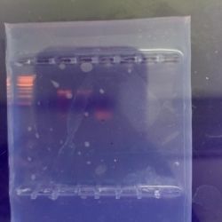
<br><center> http://i35.photobucket.com/albums/d197/corey92/9267e9ba-adcf-4c77-8ab4-1dba20b4edb1_zpsfcmqyuxz.jpg<br></center> | |||
<center>Figure1: PCR gel for bacteria found in transect 3.</center> | |||
<br>'''CONCLUSIONS & FUTURE DIRECTIONS''': | |||
After an analysis of the PCR gel for the 16S gene, researchers can conclude that DNA was present and processed because of the cuts between shorter bands of DNA where the 16S gene is present. With this presence, the use of the 16S gene can be used to identify the type of bacteria present in the transect due to the high variability of the sequence from one type of bacteria to another. | |||
The 16S gene was used to identify the tetracycline-resistant sample as Chryseobacterium which are generally raised, yellow colored colonies characterized by their rod shape. These bacteria are also generally gram negative which are inconsistent with the results of the gram staining procedure done previously to observe the bacteria. This discrepancy could be due to the lack of expertise in the area of interpretting gram stains/the gram staining was not done properly. | |||
The bacteria identified were both motile and non-motile according to literature on the bacteria which is consistent with the results of the bacteria found in the petri dishes. | |||
---- | |||
== '''LAB #6: Zebra Fish''' == | |||
'''PURPOSE''': The purpose of this lab was to learn the stages of embryonic development, compare embryonic development in different organisms, and set up an experiment to study how environmental conditions affect embryonic development in zebra fish. | |||
<br><br> | |||
'''MATERIALS & METHODS:''' | |||
<br>'''Experimental Set Up''' | |||
After perusing the current and past literature about Zebra fish experiments, an experiment was designed in order to assess the effect a given substance has on the development of a zebra fish embryo. Two groups of zebra fish embryos were prepared in two separate petri dishes with 25mLs of total solution in which the zebra fish embryos were allowed to develop. One group with 20 healthy, 24hpf (hours post fertilization) translucent embryos were transferred into a petri dish labeled “caffeine negative” along with 25mL of water. A second group was prepared with another 20 translucent embryos along with 25mL of 25mg/L caffeine solution and placed in a petri dish labeled “caffeine positive.” On the first, fourth, seventh, 11th, and 14th days of the experiment results and observations (via examination of the specimens in the petri dish under a dissection microscope) of development were recorded and dead embryos/hatchlings were removed from each set of petri dishes if present. Petri dishes were stored along with those of other researchers in plastic bins with moistened paper towels in order to preserve the humidity of the environment over the two-week period. | |||
<br>'''First Day''': | |||
On the first day after experimental set up, an observation of the developmental stage of the zebra fish embryos was conducted under a dissecting microscope for both the experimental and control groups. Water level observations and adjustments were also made in order to ensure enough water for the embryos. Dead embryos were removed if present and petri dishes were stored back in the plastic bins until the next set of observations were to take place. | |||
<br>'''Fourth Day''': | |||
On the fourth day of the experiment in addition to the removal of any dead embryos/hatchlings and developmental observations, 10mL of solution (including any waste and debris particles) was removed from the caffeine negative dish as well as 10mL of solution (including any waste and debris particles) from the caffeine positive petri dish. 25mL of water solution was then added to the caffeine negative petri dish and 25mL of 25mg/L caffeine solution was added to the caffeine positive dish. Petri dishes were then stored back in the plastic bins until the next set of observations were to take place. | |||
<br>'''Seventh Day''': | |||
On the seventh day of the experiment, developmental observations for the embyros/hatchlings were recorded for the remaining specimens as well as the addition of one drop of food (Paramecium). 10mL of water solution and caffeine solution were also added to the caffeine (+) and caffeine (-) solution petri dishes respectively. Additionally, one hatchling from each of the petri dishes (caffeine negative and caffeine positive) was removed using a dropper and placed in a tube with 1 drop of tricaine solution per mL of water a paraformaldehyde solution for closer measurement and examination. | |||
<br>'''Eleventh Day''': | |||
On the eleventh day the embryos were then assessed for size, developmental stage, motility and color, eye movement, heart rate, pectoral fin development, yolk sac size (if present), development of swim bladder and development of mouth over a two-week period. Waste particles were removed and 10mL of water solution were added to the caffeine negative dish and 10mL of caffeine to the caffeine positive petri dish. | |||
<br>'''Fourteenth Day''': | |||
On the fourteenth day final observations on morphological changes were recorded. Measurements of the length of the tail and entire hatchling as well as the measurement of the size of the eyes were recorded and the experiment was terminated. Live fish still present in the petri dishes were removed and placed in a communal environment and dead fish were discarded in a waste bowl. Petri dishes were discarded. | |||
<br><br> | |||
'''DATA & OBSERVATIONS''': | |||
<br><br> | |||
<center>http://i35.photobucket.com/albums/d197/corey92/Screen%20Shot%202015-03-18%20at%203.34.45%20PM_zpsi6jx6qza.png</center> | |||
<br><br> | |||
<center><table class="tableizer-table"> | |||
<tr class="tableizer-firstrow"><th>http://i35.photobucket.com/albums/d197/corey92/cde34682-133a-4146-91b5-90f221214410_zpsd18f6687.jpg</th><th>http://i35.photobucket.com/albums/d197/corey92/1b6e407b-8cd1-4454-b625-fbb402bcd5db_zps946e4f20.jpg</th><th>http://i35.photobucket.com/albums/d197/corey92/c4d56656-2b20-478e-97a0-6412de756f1e_zps39f68fc6.jpg</th></tr> | |||
<tr><td>'''Figure 1''': Embryo on Feb. 19</td><td>'''Figure 2''': Embryo on Feb. 20</td><td>'''Figure 3''': Hatchlings on Feb. 23</td></tr> | |||
</table> | |||
<br><br> | |||
<table class="tableizer-table"> | |||
<tr class="tableizer-firstrow"><th>http://i35.photobucket.com/albums/d197/corey92/883e278f-dacb-48bf-aa55-f01dd9be47ac_zpskxdj3hxc.jpg</th><th>http://i35.photobucket.com/albums/d197/corey92/9f532a41-f78e-4fdb-a468-83b54e0f4a96_zpsswowaazk.jpg</th></tr> | |||
<tr><td>'''Figure 4''': Hatchlings on Feb. 26 - CAF+</td><td>'''Figure 5''': Hatchlings on Feb. 26 - CAF -</td></tr> | |||
</table> | |||
<br><br> | |||
<table class="tableizer-table"> | |||
<tr class="tableizer-firstrow"><th>http://i35.photobucket.com/albums/d197/corey92/81dcd46b-676b-42f7-a77f-bf6965804674_zps41lwkypl.jpg</th><th>http://i35.photobucket.com/albums/d197/corey92/dc52ae17-ed7a-4e46-9cfc-278dd807d634_zps2gw7ysi7.jpg</th></tr> | |||
<tr><td>'''Figure 6''': Hatchlings on March 02 - CAF +</td><td>'''Figure 7''': Hatchlings on March 02 - CAF -</td></tr> | |||
</table> | |||
<br><br> | |||
<table class="tableizer-table"> | |||
<tr class="tableizer-firstrow"><th>http://i35.photobucket.com/albums/d197/corey92/9cdc67e5-758f-4243-83ce-ca98582bc7e0_zpspdellcjp.jpg</th><th>http://i35.photobucket.com/albums/d197/corey92/512d5673-4f7c-475c-a1bb-98562d61f375_zpspxx5wir4.jpg</th></tr> | |||
<tr><td>'''Figure 8''': Petri Dish on March 04 - CAF+</td><td>'''Figure 9''': Petri Dish on March 04 - CAF-</td></tr> | |||
</table> | |||
<br> | |||
{| {{table}} | |||
| align="center" style="background:#f0f0f0;"|'''Date''' | |||
| align="center" style="background:#f0f0f0;"|'''Group''' | |||
| align="center" style="background:#f0f0f0;"|'''Size''' | |||
|- | |||
| 19-Feb-15||Caffeine (+)||0.10mm | |||
|- | |||
| ||Caffeine (-)||0.10mm | |||
|- | |||
| 20-Feb-15||Caffeine (+)||0.10mm | |||
|- | |||
| ||Caffeine (-)||0.12mm | |||
|- | |||
| 23-Feb-15||Caffeine (+)||0.14mm | |||
|- | |||
| ||Caffeine (-)||0.15mm | |||
|- | |||
| 26-Feb-15||Caffeine (+)||0.16mm | |||
|- | |||
| ||Caffeine (-)||0.17mm | |||
|- | |||
| 2-Mar-15||Caffeine (+)||0.18mm | |||
|- | |||
| ||Caffeine (-)||0.19mm | |||
|- | |||
| 4-Mar-15||Caffeine (+)||0.20mm | |||
|- | |||
| ||Caffeine (-)||0.21mm | |||
|} | |||
<br> | |||
{| {{table}} | |||
| align="center" style="background:#f0f0f0;"|'''Characteristic''' | |||
| align="center" style="background:#f0f0f0;"|'''Caffeine (+) Experimental''' | |||
| align="center" style="background:#f0f0f0;"|'''Caffeine (-) Control''' | |||
|- | |||
| Eye Movement||Slowed, Inconsistent||Rapid | |||
|- | |||
| Heart Rate||76 beats/minute||64 beats/minute | |||
|- | |||
| Pectoral Fin Development||Undeveloped||Developed | |||
|- | |||
| Swim Bladder Development||Undeveloped||Developed | |||
|- | |||
| Protruding Jaw||Undeveloped||Developed | |||
|} | |||
</center> | |||
'''CONCLUSIONS AND FUTURE DIRECTIONS''': | |||
Researchers hypothesize that the presence of caffeine in solution with negatively affect the survival rate of the zebra fish embryos. Researchers also predict that if the caffeine solution is present in the petri dish, then the lifespan of the embryo and resultant zebra fish will be shortened. | |||
<br> After a two week observation of the zebra fish embryo's, researchers' observations were consistent with the aforementioned hypothesis that the zebra fish exposed to a caffeine solution would decrease the lifespan of the zebra fish as well as delay the development of motility and other characteristics of development. | |||
<br> Also, the use of the zebra fish embryos were advantageous in observing directly changes in morphology and development over the two week period which is consistent with previous literature. | |||
---- | |||
'''2.20.15''' | |||
Excellent entry. Data is clearly presented | |||
Good food web. I like that you included humans in your vertebrate list. | |||
'''SK''' | |||
---- | |||
== '''LAB #5: Invertebrates''' == | |||
'''PURPOSE''': The purpose of the current lab is to understand the importance of invertebrates as well as learn how complex systems are derived (via evolution) from simpler ones. | |||
<br> | |||
<br>'''MATERIALS & METHODS''': | |||
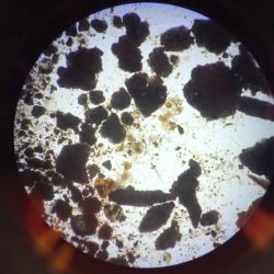
<br>''Procedure I'': An observation of the acoelomates, pseudocoelomates, and coelomates were observed via gross examination. The type of movement, as well as characteristics from microscopic examination of prepared slides of cross sections were recorded. Nematodes were also examined using a dissection microscope. | |||
<br>''Procedure II'': Students conducted a gross examination of different types of arthropods, specifically arachnida, diplopoda, chilopoda, insects, and crustacea. | |||
<br>''Procedure III'': An analysis of the Berlese funnel prepared the week prior was the third part of the lab with the purpose of collecting invertebrates from group transects. From the 25mL mixture of 50:50 ethanol/water solution in a 50mL conical tube secured to the bottom of the berlese funnel invertebrates were collected in two petri dishes, one from the top half of the conical tube and the second half from the bottom half. Microscopic examination via a dissection microscope was conducted on the two samples. | |||
<br>''Procedure IV'': A consideration of vertebrates in the transect, further discussed in the data and observations section below. | |||
<br> | |||
<br>'''DATA & OBSERVATIONS''': | |||
<br><center><table class="tableizer-table"> | |||
<tr class="tableizer-firstrow"><th>Organism (Phylum and Class)</th><th>Length in mm</th><th>Number in Sample</th><th>Description of Organism</th></tr> | |||
<tr><td>Arthropod: Ground Spider</td><td>.1mm</td><td>1</td><td>White, Eight (8) Legs</td></tr> | |||
<tr><td>Arthropod: Springtail</td><td>.2mm</td><td>2</td><td>White, Six (6) Legs</td></tr> | |||
<tr><td>Arthropod: Centipede</td><td>.7mm</td><td>1</td><td>White, >20 Legs</td></tr> | |||
</table> | |||
<br>'''''Table 1''''': Summary of invertebrates identified in transect #3 at American University. Identification was done via the use of key displaying common soil invertebrates.</center> | |||
<br> | |||
<center><table class="tableizer-table"> | |||
<tr class="tableizer-firstrow"><th>http://i35.photobucket.com/albums/d197/corey92/addc84a1-e69e-491c-8389-37f2f0ab8a4e_zpseb38b6ce.jpg</th><th>http://i35.photobucket.com/albums/d197/corey92/73f2fc23-9552-4ad3-b26a-6108c07b8ed0_zpsb1add99a.jpg</th><th>http://i35.photobucket.com/albums/d197/corey92/9ece5650-af61-43a2-ac0c-c8165614ac4b_zps7773b843.jpg</th></tr> | |||
<tr><td>'''Figure 1''': 10X magnification of Garden Spider</td><td>'''Figure 2''': 10X magnification of Spring tail</td><td>'''Figure 3''': 10X magnification of Centipede</td></tr> | |||
</table></center> | |||
<br> | |||
<br><center><table class="tableizer-table"> | |||
<tr class="tableizer-firstrow"><th>Organism </th><th>Classsification</th><th>Abiotic Features (& Benefits)</th><th>Biotic Features (& Benefits)</th></tr> | |||
<tr><td>Human</td><td>Chordata, Mammalia, Primates, Hominidae, Homonini, Homo Sapiens </td><td>Water (Food)</td><td>Trees (Shade, Oxygen)</td></tr> | |||
<tr><td>Raccoon</td><td>Chordata, Mammalia, Carnivora, Procyonidae, Prycon, Prycon lotor </td><td>Water (Food), Trash (Food)</td><td>Trees (Shelter and Oxygen), Insects (Food), Plants (Food)</td></tr> | |||
<tr><td>Squirrel</td><td>Chordata, Mammalia, Rodentia, Sciuramorpha, Sciuridae, Sciurus </td><td>Water (Food), Seeds (Food), Nuts (Food)</td><td>Trees (Shelter & Oxygen)</td></tr> | |||
<tr><td>Blue Jay</td><td>Chordata, Aves, Passeriformes, Corvidae, Cyanocitta, Cyanocitta cristata</td><td>Water (Food), Seeds (Food), Dead Foliage (Nests & Shelter)</td><td>Trees (Shelter & Oxygen), Insects (Food)</td></tr> | |||
<tr><td>Woodpecker </td><td>Chordata, Aves, Piciformes, Pici, Picides, Picidae, Picinae</td><td>Water (Food), Seeds (Food), Dead leaves (nests) </td><td>Trees (Shelter and Oxygen), Insects (Food)</td></tr> | |||
</table> | |||
<br>'''''Table 2''''': Summary of vertebrates identified in transect #3. | |||
<br> | |||
<br>http://i35.photobucket.com/albums/d197/corey92/IMG_8609copy_zps2d16baa2.jpg | |||
Figure 4: Food web diagram</center> | |||
<br>'''CONCLUSIONS & FUTURE DIRECTIONS''': | |||
Many of the invertebrates observed in lab, relative to their movement, had no direct form of predictable motion. Their modus operandi was a writhing motion with no predictable pattern of motion which makes sense when considering the lack of vertebrae and more formalized body structure. | |||
<br> | |||
Although limited in number, the invertebrates identified in transect #3 did well to provide us with insight into the diversity of life within the transect. The identification of three different types of invertebrates should and does not limit the possibility of many other organisms being present. In addition to the invertebrates identified in the transect, vertebrates or chordata were also identified. Relative to the idea of community, these organisms are that which form the community within the transect via direct and indirect interaction. The concept of carrying capacity is illustrated via the limitations of the resources of the transect that can support a particular number of individuals, and lastly, the concept of trophic levels or the space an organism occupies in a food chain. Vertebrates which are in most cases larger than the invertebrates most likely feed on the smaller invertebrates within the transect. An example of this is a bird eating a spider or a centipede; however, the centipede and spider will be in a different trophic level than the birds and humans in the transect due to the nature of their feeding on plant matter, etc. | |||
*'''[[User:Corey J Salas|Corey J Salas]] 00:17, 19 February 2015 (EST)''': | |||
---- | |||
'''2.20.15''' | |||
Excellent notebook entry. | |||
'''SK''' | |||
== '''LAB #4: Plantae and Fungi''' == | |||
'''PURPOSE''': The general purpose of this lab is to understand and appreciate the characteristics, functions, importance and most especially the diversity of plants and fungi. | |||
<br> | |||
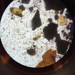
<br>'''MATERIALS & METHODS''': The current laboratory assignment was divided into six (6) sections wherein different materials and methods were used. In the first part of the experiment ziploc bags were used to obtain a leaf litter sample from the group's assigned lab transect consisting of dead leave and plant matter as well as a small sample of the top soil. Secondarily, five (5) plant samples representative of the transect were collected for observation in the lab. The second part of the lab was a comparison of vascularity in Bryophytes [Moss} (rhizoids) vs. Angiosperms (xylem and phloem). A comparison of the moss, ''Mnium'' height was compared to the height of the lily plant as well as an identification of the xylem and phloem laters of prepared cross section slide of a lily stem. The third part of the experiment used microscopy to identify the different components of leaf anatomy. The structures being identified were features such as stomata, guard cells, mesophyll, parenchyma, palisade mesophyll, etc. In the fourth part of the experiment the reproductive components of plants were observed. A particular focus was given to the gametophyte (haploid) vs. sporophyte (diploid) stages of the lifecycle. A lily flower was then dissected and features such as the anther, stigma, style, etc. were observed via a gross examination as well as under the microscope in order to gain a clearer understanding of and appreciation for plants. | |||
<br> | |||
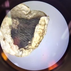
<br>Part five of the experiment requires a microscopic examination and identification of 3 different types of fungi; zygomycota, basidiomycota, and ascomycota (Figures 1-3). Part six of the laboratory was a preparation of a Burlese Funnel to collect invertebrates from group transects. To prepare the funnel, a 25mL mixture of 50:50 ethanol/water solution was placed in a 50mL conical tube which was secured to the bottom of the burlese funnel, a screen was placed in the bottom (inner) of the funnel and secured with tape. The leaf litter sample was then placed in the top of the funnel, secured to a ring stand and placed in a foil tent with a 40-watt lamp approximately 1-2 inches from the top of the leaf litter. The observations of the burlese funnel will be recorded next week. | |||
<br> | |||
<br>'''DATA & OBSERVATIONS''': | |||
<br><center><table class="tableizer-table"> | |||
<tr class="tableizer-firstrow"><th>Microscope #1</th><th>Microscope #2</th><th>Microscope #3</th></tr> | |||
<tr><td>http://i35.photobucket.com/albums/d197/corey92/5dbd71ea-b31a-4f1c-a72c-7da78976ad95_zps655ee218.jpg</td><td>http://i35.photobucket.com/albums/d197/corey92/8c924513-cd70-484c-bec3-53d9c7d668cd_zps30c2a8f8.jpg</td><td>http://i35.photobucket.com/albums/d197/corey92/4018237c-242f-4107-99b7-30261997f069_zps5eebf3f2.jpg</td></tr> | |||
<tr><td>'''''Figure 1''''': Basidiomycota (Mushroom)</td><td>'''''Figure 2''''': Zygomycota (''Rhizopus Nigricans'')</td><td>'''''Figure 3''''': Ascomycota (''Rhizopus stolonifer zygospore'')</td></tr> | |||
</table></center> | |||
<br>In an observation of the different examples of fungi above it is important to note the specialized sporangia and their function as the receptacle in which asexual spores for reproduction are formed. The three different types of fungi observed in microscopes 1-3 were classified as Basidiomycota, zygomycota, and ascomycota respectively. | |||
<center><table class="tableizer-table"> | |||
<tr class="tableizer-firstrow"><th>http://i35.photobucket.com/albums/d197/corey92/f654163b-62f4-47ca-8718-55598dd0fab5_zps3fd1ad56.jpg</th><th>http://i35.photobucket.com/albums/d197/corey92/3451b2ca-fda0-479e-8954-8f923ae5d814_zps22807297.jpg</th></tr> | |||
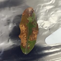
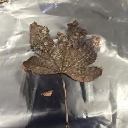
<tr><td>'''''Figure 4''''': Leaf sample #1 from transect 3</td><td>'''''Figure 5''''': Leaf sample #2 from transect 3</td></tr> | |||
</table></center> | |||
<center><table class="tableizer-table"> | |||
<tr class="tableizer-firstrow"><th>http://i35.photobucket.com/albums/d197/corey92/b8b21ede-51f2-4a49-92e0-0aa085c45cd1_zps178401fc.jpg </th><th> http://i35.photobucket.com/albums/d197/corey92/e249ec1e-8ee1-4f77-8a63-58f4b0c9582a_zpsbb0eec00.jpg</th><th>http://i35.photobucket.com/albums/d197/corey92/f5d8773e-f1e5-404f-8d11-ecf6416f5219_zps0aea992c.jpg</th></tr> | |||
<tr><td>'''''Figure 6''''': Leaf sample #3 from transect 3</td><td>'''''Figure 7''''': Leaf sample #4 from transect 3</td><td>'''''Figure 8''''': Leaf sample #5 from transect 3</td></tr> | |||
</table></center> | |||
<center>'''''Figures 4-8''''': images showing the different leaf samples obtained from transect #3.</center> | |||
<br> The samples obtained from the transect were relatively small in size (see Table 1 below) and were scattered for the most part on the floor of the area; this leaf placement is most likely due to the seasonal changes in plants that are brought on by the winter months. So too, the table below describes the vascularization of each of the plants from out transect as well as other feature details. | |||
<br><br> | |||
<table> | |||
<tr> | |||
<td>'''Transect Sample Plants'''</td> | |||
<td>'''Location and # in Transect'''</td> | |||
<td>'''Description (Size & Shape)''' </td> | |||
<td>'''Vascularization'''</td> | |||
<td>'''Specialized Structures'''</td> | |||
<td>'''Mechanism of Reproduction''' </td> | |||
</tr> | |||
<tr> | |||
<td>#1</td> | |||
<td>Shrub (~500) </td> | |||
<td>Dying, curling (5.0cm x 3.0cm)</td> | |||
<td>Branched veins</td> | |||
<td></td> | |||
<td>Alternation of Generations</td> | |||
</tr> | |||
<tr> | |||
<td>#2</td> | |||
<td>Ground Level (~500)</td> | |||
<td>Green, spotted with brown, wrinkled (6.0cm x 2.5cm)</td> | |||
<td>Branched Veins</td> | |||
<td></td> | |||
<td>Alternation of Generations</td> | |||
</tr> | |||
<tr> | |||
<td>#3</td> | |||
<td>Ground Level (~500)</td> | |||
<td>Grayish brown, curling, dying, cracked surface, vasularity visible (4.0cm x 5.5cm) </td> | |||
<td>Branched Veins</td> | |||
<td></td> | |||
<td>Alternation of Generations</td> | |||
</tr> | |||
<tr> | |||
<td>#4</td> | |||
<td>Ground Level (~500)</td> | |||
<td>Porous, dead, brittle with rigid edges (4.5cm x 2.0cm)</td> | |||
<td>Branched Veins</td> | |||
<td>Vascular net structure</td> | |||
<td>Alternation of Generations</td> | |||
</tr> | |||
<tr> | |||
<td>#5</td> | |||
<td>Tree Level (~75) </td> | |||
<td>Green, alive, intact, multiple leaves on sample branch, smooth surface, pointed edges (4.0cm x 1.5cm) </td> | |||
<td>Branched Veins</td> | |||
<td></td> | |||
<td>Alternation of Generations</td> | |||
</tr> | |||
</table> | |||
<br>'''CONCLUSIONS & FUTURE DIRECTIONS''': The diversity of plants, fungi, and animals can be seen right in front of us. The small area in which we have obtained samples since the beginning of the semester has provided us with a multitude of organisms, concepts, and ideas to observe and relate back to our study of biology. One must not observe all the diversity that exists on this planet in order to study and understand the concept of biodiversity. The main goals of this lab was attained in the observations made in the samples above as well as the preparation of the leaf litter for observation next week. | |||
*'''[[User:Corey J Salas|Corey J Salas]] 01:18, 10 February 2015 (EST)''': | |||
---- | |||
'''2.10.15''' Excellent lab book entry containing all relevant data and methods. The low magnification image of gram stained slides does't really serve a purpose. '''SK''' | |||
== '''LAB #3: Microbiology and Identifying Bacteria with DNA Sequences''' == | |||
'''PURPOSE''': | |||
<br>The general purpose of this lab was to understand the characteristics of bacteria such as shape, size, motility, shape and arrangement as well as to observe antibiotic resistance using the bacteria found in our hay infusion culture and to use DNA sequences in order to identify species. | |||
<br>'''MATERIALS & METHODS''': | |||
<br>The first part of the experiment was to quantify and observe microorganisms that had grown in our bacterial cultures. The materials used in this lab were nutrient rich agar plates in two sets, one set with +TET and one set without prepared with bacteria during the lab section the previous week. Microscopy was used to observe bacterial culture (1 week growth time). | |||
<br><br>In the second part of the experiment, in order to identify antibiotic resistance, a gram staining procedure was used. In order to gram stain the bacteria solutions of crystal violet, Gram's iodine, 95% alcohol and water were used. The prepared slides were then observed at 40X and 100X (oil immersion) lenses. | |||
<br><br>In the third part of the lab, a PCR for 16S sequencing was set up using one of the four different bacterial colonies in the hay infusion culture. A sample of the bacteria in the colony was transferred into a 100μL sterile tube, incubated at 100°C for 10 minutes, centrifuged for 5 minutes at 13,400rpm, mixed with a primer and then placed in the PCR machine. Results of the PCR will be analyzed next week on an agrose gel in order to properly identify the bacteria present in the hay infusion culture. | |||
<br>'''DATA & OBSERVATIONS''': | |||
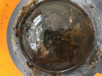
<br> Upon observation and contact with the hay infusion culture, it is notable that the smell had changed from the initial preparation approximately 14 days prior. The smell of putrefaction had disappeared from the jar as well as a white film that had formed the week prior in the top portion of the jar. The water level in the jar had also decreased approximately 1 inch from the level the week prior. | |||
<br>In part one of the experiment in order to quantify and identify organisms the following table summarizes the observations: | |||
<center>'''Table 1: 100-fold Serial Dilutions Results''' | |||
<br>http://i35.photobucket.com/albums/d197/corey92/9204068f-5fca-449d-bb11-ba6a23f2e621_zps818c21dc.png</center> | |||
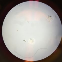
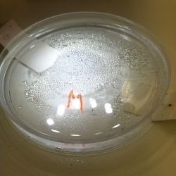
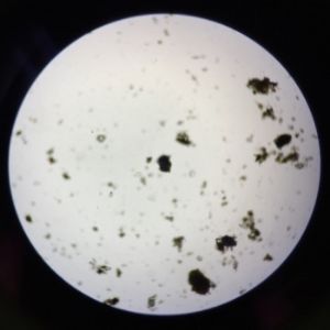
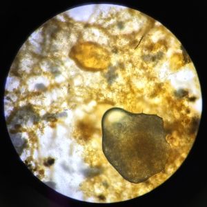
<br> Observations of the agar plates, both TET rich and deficient, had different types of bacteria present. Some colonies were round in shape, others were more differentiated although still circular. Some of the bacterial colonies were yellow in color and others were white with very clear differentiation between. Most of the colonies were raised as opposed to flat and glossy in color. A wet mount of four of the bacterial colonies were observed at 100x magnification before staining; however, photos of the stained bacteria produce a more quality image and representation of the shape, size, and motility of the bacteria and are therefore used in this lab entry. A photo of the colonies can be found below: | |||
<br><center>http://i35.photobucket.com/albums/d197/corey92/89ca2d2f-bdff-4d91-a437-d872fa639342_zps2c9fbd9a.jpg | |||
'''Figure1''': All AGAR plates with bacteria from hay infusion. Top Row: AGAR -TET Bottom Row: AGAR +TET | |||
<br> | |||
<br>http://i35.photobucket.com/albums/d197/corey92/0445a37a-630a-4297-b18f-d5fbfd44a513_zps6e363789.jpg | |||
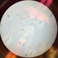
'''Figure2''': Image of AGAR plates with colonies for observations. Colonies </center> | |||
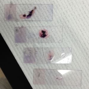
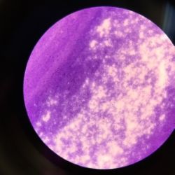
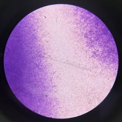
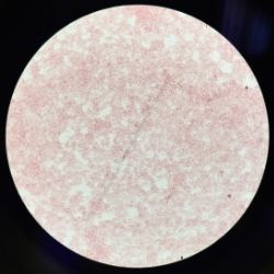
<br>In part 2 of the experiment, an assessment of antibiotic resistance was done via gram staining. Pictures of the gram stain are below: | |||
<br><center>http://i35.photobucket.com/albums/d197/corey92/fa2c0f98-785b-44ec-9193-2374c0000306_zps1a9c8b69.jpg | |||
'''Figure 3''': Image of gram stain slides #1-4 | |||
<br>http://i35.photobucket.com/albums/d197/corey92/291c2dee-3e36-4d7d-b0a2-ba09b4f3b728_zps4efd7361.jpg | |||
<br> | |||
'''Figure 4''': Slide #1 showing gram positive bacteria sample from TET+ plate (100X) | |||
<br>http://i35.photobucket.com/albums/d197/corey92/60c28e1a-94c9-40d3-ac5d-9f360b9ebb53_zpsd4b74a75.jpg | |||
<br> | |||
'''Figure 5''': Slide #2 showing gram positive bacteria sample from TET+ plate (100X) | |||
<br>http://i35.photobucket.com/albums/d197/corey92/65c9e50a-89e2-460b-89a7-88daf4e40af0_zpsfbfd09c8.jpg | |||
<br> | |||
'''Figure 6''': Slide #3 showing gram negative bacteria sample from AGAR -TET (100x) | |||
<br> http://i35.photobucket.com/albums/d197/corey92/6d15fba0-5390-47ad-8533-1225bec66ce0_zpse1b58d1c.jpg | |||
<br> | |||
'''Figure 7''': Slide #4 showing gram negative bacteria sample from AGAR -TET (100X) </center> | |||
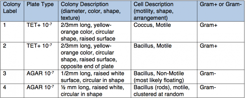
A summary of observations of bacteria are as follows: | |||
<center>'''Table 2:Bacterial Characterization''' | |||
<br>http://i35.photobucket.com/albums/d197/corey92/ScreenShot2015-02-05at120636AM_zps323b4c0d.png</center> | |||
<br>'''CONCLUSIONS & FUTURE DIRECTIONS''': | |||
<br>Gram positive bacteria are antibiotic resistant based on the observation that only the gram positive bacteria existed on the Agar + TET plates. In terms of the observations made on the plates, observations are in line with the mechanisms of action as discussed in 2001 by Ian Chopra and Marilyn Roberts, "Tetracyclines traverse the outer membrane of gram-negative enteric bacteria through the OmpF and OmpC porin channels, as positively charged cation (probably magnesium)-tetracycline coordination complexes (44, 263). The cationic metal ion-antibiotic complex is attracted by the Donnan potential across the outer membrane, leading to accumulation in the periplasm, where the metal ion-tetracycline complex probably dissociates to liberate uncharged tetracycline, a weakly lipophilic molecule able to diffuse through the lipid bilayer regions of the inner (cytoplasmic) membrane." Also notable was the absence of any organisms classified as archaea in the plates which makes sense because archaea generally thrive in areas of extreme environmental conditions. | |||
*'''[[User:Corey J Salas|Corey J Salas]] 22:50, 4 February 2015 (EST)''': | |||
---- | |||
'''2.4.15''' Excellent lab book entry. Nice detailed description of Hay Infusion and particular protists found within. Logical and well organized. | |||
Beautiful diagram of serial dilutions! '''SK''' | |||
== '''LAB #2: Identifying Algae and Protists''' == | |||
'''PURPOSE''': | |||
<br> The general purpose of this lab was to practice using a dichotomous key to identify unknown algae and protists from our transect at American University as well as gaining a clearer understanding of the characteristics of algae and protists. | |||
<br>'''MATERIALS & METHODS''': | |||
<br> This lab was divided into three main parts. The first was to assist students in using a dichotomous key, the second was to identify life at the microbiological level in the hay infusion culture prepared the previous week, and third to prepare and plate a serial dilution. In the first part of the lab, students were instructed to go around the room and observe different microscopic slides and use the dichotomous key to attempt to align their observations with the organism deduced using the key. Students were then instructed to prepare a wet mount of known organisms and observe them with the microscope at 4x and 10x magnifications. | |||
<br> | |||
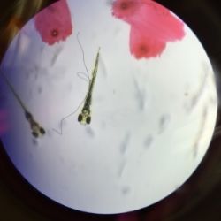
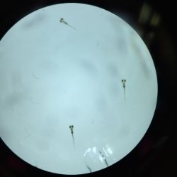
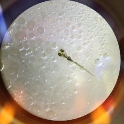
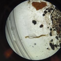
<br> In the second part of the lab, samples were taken from the hay infusion culture from the bottom, middle, and top sections arbitrarily assigned by the group members. Wet mounts were prepared and a determination of which protists or algae was undertaken using the dichotomous key. Figures 1-4 below show some of the external, internal and microscopic observations. | |||
<br> | |||
<br> The third part of the lab required students to create a serial dilution with a sample of the hay infusion culture and 10mLs of sterile broth. Four test tubes labeled 10^2, 10^4, 10^6, 10^8 with approximately 10mLs of sterile brother were used in the dilution. 100µl of the culture were added to the 10mLs of broth labeled 10^2, 10µl of the broth from the tube labeled 10^2 were then added to the tube labeled 10^6, 100µl of the broth from the tube labeled 10^6 were then added to the tube labeled 10^8 which completed the serial dilution preparation. These dilutions were then plated on the nutrient rich agar plates in two sets, one set with +TET and one set without. See the serial dilution diagram below (''Figure 5''). | |||
<br>'''DATA & OBSERVATIONS''': | |||
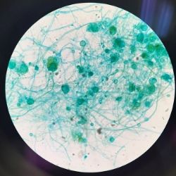
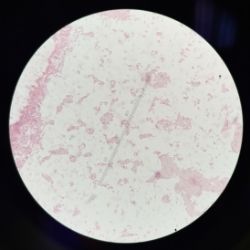
<br> The second portion of the lab yielded the most interesting results as organisms from the group hay infusion culture were being observed. All organisms observed appeared to be motile and were in both the algae and protazoan lineages. None of the organisms were more than 200µl in size and were not visible to the naked eye; therefore an assessment of their loitering near plant matter in the infusion culture is difficult . Observed in the samples placed on the wet mounts were the following: | |||
<br><center> ''Top'': Chlamydomonas, Euplotes sp. ''Middle'': Busaria Truncatella & Paramecium Aurelia ''Bottom'': Paramecium Bursaria & Vorticella </center> | |||
<br> Upon initial contact with the hay infusion culture, it is notable that the smell had changed from the initial preparation approximately 7 days prior. There was a smell of putrefaction exuding from the jar as well as a white film that had formed in the top portion of the jar - the group assumes this is some type of mold. | |||
<center>http://i35.photobucket.com/albums/d197/corey92/80c06dfa-4e75-43d9-96ca-57d17874c5fc_zpsea1eab04.jpg http://i35.photobucket.com/albums/d197/corey92/54c8cfee-37db-42bc-9a27-2017949fbc98_zps91100938.jpg | |||
<br><br> http://i35.photobucket.com/albums/d197/corey92/1d54e7bc-d75a-4d97-9a08-8281a6d174a3_zps3e6e7af0.jpg http://i35.photobucket.com/albums/d197/corey92/34c8d0aa-8417-4a12-9c91-9066bac5ee23_zps698186af.jpg | |||
Figures 1-4: Hay infusion culture exterior and microscopic observations.</center> | |||
<br> | |||
<br> | |||
<center>http://i35.photobucket.com/albums/d197/corey92/8d1aa4f3-65ca-434a-9914-17817c5ab697_zpsf0e63d12.png | |||
<br>Figure 5: Serial dilution of hay infusion cultures to agar plates.</center> | |||
<br>'''CONCLUSIONS & FUTURE DIRECTIONS''': | |||
<br> Due to the biological nature of our study this semester, it is important to relate what we observe back to what we know about our definition of life. Thinking about the organisms observed in the hay culture, chlamydomonas serve well to bridge the gap between how we define life and how we observe the characteristics of that definition. Chlamydomonas has cells, uses energy (photosynthesis), grow and can reproduce - these are all characteristics that define life as well as characteristics of chlamydomonas itself. Thinking forward about how this culture might grow in the future, depending on environmental conditions, the infusion culture might grow more organisms or could very well kill them off due to lack of resources, it really all depends. | |||
*'''[[User:Corey J Salas|Corey J Salas]] 17:16, 28 January 2015 (EST)''': | |||
'''1.27.15''' | '''1.27.15''' | ||
Excellent first lab book entry. Well structured and organized. Nice photo. Transect is 20 ft by 20 ft there is a typo in the manual. | Excellent first lab book entry. Well structured and organized. Nice photo. Transect is 20 ft by 20 ft there is a typo in the manual. | ||
| Line 27: | Line 376: | ||
<br>Based on the observations of the transect and the sample collected and prepared in the hay infusion culture, it is advantageous to say that there is much biodiversity present in and around the American University campus. It is also important to note that the changing biotic and abiotic features of the transect will be of particular interest to observe as the semester progresses. | <br>Based on the observations of the transect and the sample collected and prepared in the hay infusion culture, it is advantageous to say that there is much biodiversity present in and around the American University campus. It is also important to note that the changing biotic and abiotic features of the transect will be of particular interest to observe as the semester progresses. | ||
--[[User:Corey J Salas|Corey J Salas]] 22:54, 26 January 2015 (EST) | <br>--[[User:Corey J Salas|Corey J Salas]] 22:54, 26 January 2015 (EST) | ||
Latest revision as of 20:52, 25 March 2015
Bacterial PCR Data
PURPOSE: The purpose of the PCR lab was to identify the type of bacteria present in the assigned transect by isolation and amplification of a single gene 16S.
MATERIALS AND METHODS:
PCR for 16S sequencing was set up using one of the four different bacterial colonies in the hay infusion culture. A sample of the bacteria in the colony was transferred into a 100μL sterile tube, incubated at 100°C for 10 minutes, centrifuged for 5 minutes at 13,400rpm, mixed with a primer and then placed in the PCR machine. Results of the PCR were sequenced using GeneWiz and identified using the NIH gene sequence blast.
DATA & OBSERVATIONS:
MB49 - NNNNNNNNNNNNNNNNANNNTGCAGCCGAGCGGTATTTGTCCTTCGGGACAGAGAGAGCGGCGTACGGGTGCGGAACACG TGTGCAACCTACCTTTATCAGGGGGATAGCCTTTCGAAAGGAAGATTAATACCCCATAATATAAGTCAAGGCATCTTGAT TTATTGAAAACTCCGGTGGATAGAGATGGGCACGCGCAAGATTAGATAGTTGGTAGGGTAACGGCCTACCAAGTCAATGA TCTTTAGGGGGCCTGAGAGGGTGATCCCCCACACTGGTACTGAGACACGGACCAGACTCCTACGGGAGGCAGCAGTGAGG AATATTGGACAATGGGTGAGAGCCTGATCCAGCCATCCCGCGTGAAGGACGACGGCCCTATGGGTTGTAAACTTCTTTTG TATAGGGATAAACCTACTCTCGTGAGAGTANCTGAAGGTACTATACGAATAAGCACCGGCTAACTCCGTGCCAGCAGCCG CGGTAATACGGAGGGTGCAAGCGTTATCCGGATTTATTGGGTTTAAAGGGTCCGTAGGCGGGCTTGTAAGTCAGTGGTGA AATCTCATAGCTTAACTATGAAACTGCCATTGATACTGCAGGTCTTGAGTAAAGTAGAAGTGGCTGGAATAAGTAGTGTA GCGGTGAAATGCATAGATATTACTTANAACACCAATTGCGAAGGCAGGTCACTATGTTTTAACTGACGCTGATGGACGAA AGCGTGGGGAGCGAACAGGATTANATACCCTGGTAGTCCACGCCGTAAACGATGCTNACTCGTTTTTGGGCTTTCGGGTT CAGAGACTAAGCGAAAGTGATAAGTTAGCCACCTGGGGAGTACGTTCGCAAGAATGAAACTCAAAGGAATTGACGGGGGC CCGCACAANCGGTNNTTATGTGGNTTAATTCGATGATANNCGAGGAACCTTANCAAAGGCTNAAATGGGAATTGACAGGN TTANAAAATAGACTTTTCTTCNNACNATTTTCAAGNTGCTGCATGGNNGTCNNCAGCTCGTGCCNTGAGTGTNGNTAAGT CCTGCAACNANCNCAACCCNGNNNNTANNTNNCATNNTTCAGTTNGGGANNNNTAGNNNN
Chryseobacterium sp. LDVH 3 16S ribosomal RNA gene, partial sequence
MB50 - NNNNNNNNNNNNNNNNNNNNNANNNNTGCAGCCGAGCGGTATTGTTTCTTCGGAAATGAGAGAGCGGCGTACGGGTGCGG ANCNNNTGTGCAACCTGCCTTTATCTGGGGGATAGCCTTTCGAAAGGGAGATTAATACCCCATAATATATTAAGTGGCAT CACTTGATATTGAAAACTCCGGTGGATAGAGATGGGCACGCGCAAGATTAGATAGTTGGTGAGGTAACGGCTCACCAAGT CTACGATCTTTAGGGGGCCTGAGAGGGTGATCCCCCACACTGGTACTGAGACACGGACCAGACTCCTACGGGAGGCAGCA GTGAGGAATATTGGACAATGGGTGAGAGCCTGATCCAGCCATCCCGCGTGAAGGACGACGGCCCTATGGGTTGTAAACTT CTTTTGTATAGGGATAAACCTACTCTCGTGAGAGTAGCTGAAGGTACTATACGAATAAGCACCGGCTAACTCCGTGCCAG CAGCCGCGGTAATACGGAGGGTGCAAGCGTTATCCGGATTTATTGGGTTTAAAAGGGTCCGTANGCGGATCTGTAAGTCA GTGGTGAAATCTCACAGCTTAACTGTGAAAACTGCCATTGATACTGCAGGTCTTGAGTGTTGTTGAAGTANCTGGAATAA GTAGTGTANCGGTGAAATGGCNTAGATATTACTTAGAAACACCAATTGCNAAGGCTNGTTACTAANCAACAACTGACNCT GATGGACGAAANCGTGGNGGAGCGAACAGGATTANATACCCCTGGNAN
Chryseobacterium sp. StRB028 gene for 16S rRNA, partial sequence
CONCLUSIONS & FUTURE DIRECTIONS:
After an analysis of the PCR gel for the 16S gene, researchers can conclude that DNA was present and processed because of the cuts between shorter bands of DNA where the 16S gene is present. With this presence, the use of the 16S gene can be used to identify the type of bacteria present in the transect due to the high variability of the sequence from one type of bacteria to another.
The 16S gene was used to identify the tetracycline-resistant sample as Chryseobacterium which are generally raised, yellow colored colonies characterized by their rod shape. These bacteria are also generally gram negative which are inconsistent with the results of the gram staining procedure done previously to observe the bacteria. This discrepancy could be due to the lack of expertise in the area of interpretting gram stains/the gram staining was not done properly.
The bacteria identified were both motile and non-motile according to literature on the bacteria which is consistent with the results of the bacteria found in the petri dishes.
LAB #6: Zebra Fish
PURPOSE: The purpose of this lab was to learn the stages of embryonic development, compare embryonic development in different organisms, and set up an experiment to study how environmental conditions affect embryonic development in zebra fish.
MATERIALS & METHODS:
Experimental Set Up
After perusing the current and past literature about Zebra fish experiments, an experiment was designed in order to assess the effect a given substance has on the development of a zebra fish embryo. Two groups of zebra fish embryos were prepared in two separate petri dishes with 25mLs of total solution in which the zebra fish embryos were allowed to develop. One group with 20 healthy, 24hpf (hours post fertilization) translucent embryos were transferred into a petri dish labeled “caffeine negative” along with 25mL of water. A second group was prepared with another 20 translucent embryos along with 25mL of 25mg/L caffeine solution and placed in a petri dish labeled “caffeine positive.” On the first, fourth, seventh, 11th, and 14th days of the experiment results and observations (via examination of the specimens in the petri dish under a dissection microscope) of development were recorded and dead embryos/hatchlings were removed from each set of petri dishes if present. Petri dishes were stored along with those of other researchers in plastic bins with moistened paper towels in order to preserve the humidity of the environment over the two-week period.
First Day:
On the first day after experimental set up, an observation of the developmental stage of the zebra fish embryos was conducted under a dissecting microscope for both the experimental and control groups. Water level observations and adjustments were also made in order to ensure enough water for the embryos. Dead embryos were removed if present and petri dishes were stored back in the plastic bins until the next set of observations were to take place.
Fourth Day:
On the fourth day of the experiment in addition to the removal of any dead embryos/hatchlings and developmental observations, 10mL of solution (including any waste and debris particles) was removed from the caffeine negative dish as well as 10mL of solution (including any waste and debris particles) from the caffeine positive petri dish. 25mL of water solution was then added to the caffeine negative petri dish and 25mL of 25mg/L caffeine solution was added to the caffeine positive dish. Petri dishes were then stored back in the plastic bins until the next set of observations were to take place.
Seventh Day:
On the seventh day of the experiment, developmental observations for the embyros/hatchlings were recorded for the remaining specimens as well as the addition of one drop of food (Paramecium). 10mL of water solution and caffeine solution were also added to the caffeine (+) and caffeine (-) solution petri dishes respectively. Additionally, one hatchling from each of the petri dishes (caffeine negative and caffeine positive) was removed using a dropper and placed in a tube with 1 drop of tricaine solution per mL of water a paraformaldehyde solution for closer measurement and examination.
Eleventh Day:
On the eleventh day the embryos were then assessed for size, developmental stage, motility and color, eye movement, heart rate, pectoral fin development, yolk sac size (if present), development of swim bladder and development of mouth over a two-week period. Waste particles were removed and 10mL of water solution were added to the caffeine negative dish and 10mL of caffeine to the caffeine positive petri dish.
Fourteenth Day:
On the fourteenth day final observations on morphological changes were recorded. Measurements of the length of the tail and entire hatchling as well as the measurement of the size of the eyes were recorded and the experiment was terminated. Live fish still present in the petri dishes were removed and placed in a communal environment and dead fish were discarded in a waste bowl. Petri dishes were discarded.
DATA & OBSERVATIONS:
| http://i35.photobucket.com/albums/d197/corey92/cde34682-133a-4146-91b5-90f221214410_zpsd18f6687.jpg | http://i35.photobucket.com/albums/d197/corey92/1b6e407b-8cd1-4454-b625-fbb402bcd5db_zps946e4f20.jpg | http://i35.photobucket.com/albums/d197/corey92/c4d56656-2b20-478e-97a0-6412de756f1e_zps39f68fc6.jpg |
|---|---|---|
| Figure 1: Embryo on Feb. 19 | Figure 2: Embryo on Feb. 20 | Figure 3: Hatchlings on Feb. 23 |
| http://i35.photobucket.com/albums/d197/corey92/883e278f-dacb-48bf-aa55-f01dd9be47ac_zpskxdj3hxc.jpg | http://i35.photobucket.com/albums/d197/corey92/9f532a41-f78e-4fdb-a468-83b54e0f4a96_zpsswowaazk.jpg |
|---|---|
| Figure 4: Hatchlings on Feb. 26 - CAF+ | Figure 5: Hatchlings on Feb. 26 - CAF - |
| http://i35.photobucket.com/albums/d197/corey92/81dcd46b-676b-42f7-a77f-bf6965804674_zps41lwkypl.jpg | http://i35.photobucket.com/albums/d197/corey92/dc52ae17-ed7a-4e46-9cfc-278dd807d634_zps2gw7ysi7.jpg |
|---|---|
| Figure 6: Hatchlings on March 02 - CAF + | Figure 7: Hatchlings on March 02 - CAF - |
| http://i35.photobucket.com/albums/d197/corey92/9cdc67e5-758f-4243-83ce-ca98582bc7e0_zpspdellcjp.jpg | http://i35.photobucket.com/albums/d197/corey92/512d5673-4f7c-475c-a1bb-98562d61f375_zpspxx5wir4.jpg |
|---|---|
| Figure 8: Petri Dish on March 04 - CAF+ | Figure 9: Petri Dish on March 04 - CAF- |
| Date | Group | Size |
| 19-Feb-15 | Caffeine (+) | 0.10mm |
| Caffeine (-) | 0.10mm | |
| 20-Feb-15 | Caffeine (+) | 0.10mm |
| Caffeine (-) | 0.12mm | |
| 23-Feb-15 | Caffeine (+) | 0.14mm |
| Caffeine (-) | 0.15mm | |
| 26-Feb-15 | Caffeine (+) | 0.16mm |
| Caffeine (-) | 0.17mm | |
| 2-Mar-15 | Caffeine (+) | 0.18mm |
| Caffeine (-) | 0.19mm | |
| 4-Mar-15 | Caffeine (+) | 0.20mm |
| Caffeine (-) | 0.21mm |
| Characteristic | Caffeine (+) Experimental | Caffeine (-) Control |
| Eye Movement | Slowed, Inconsistent | Rapid |
| Heart Rate | 76 beats/minute | 64 beats/minute |
| Pectoral Fin Development | Undeveloped | Developed |
| Swim Bladder Development | Undeveloped | Developed |
| Protruding Jaw | Undeveloped | Developed |
CONCLUSIONS AND FUTURE DIRECTIONS:
Researchers hypothesize that the presence of caffeine in solution with negatively affect the survival rate of the zebra fish embryos. Researchers also predict that if the caffeine solution is present in the petri dish, then the lifespan of the embryo and resultant zebra fish will be shortened.
After a two week observation of the zebra fish embryo's, researchers' observations were consistent with the aforementioned hypothesis that the zebra fish exposed to a caffeine solution would decrease the lifespan of the zebra fish as well as delay the development of motility and other characteristics of development.
Also, the use of the zebra fish embryos were advantageous in observing directly changes in morphology and development over the two week period which is consistent with previous literature.
2.20.15 Excellent entry. Data is clearly presented Good food web. I like that you included humans in your vertebrate list. SK
LAB #5: Invertebrates
PURPOSE: The purpose of the current lab is to understand the importance of invertebrates as well as learn how complex systems are derived (via evolution) from simpler ones.
MATERIALS & METHODS:
Procedure I: An observation of the acoelomates, pseudocoelomates, and coelomates were observed via gross examination. The type of movement, as well as characteristics from microscopic examination of prepared slides of cross sections were recorded. Nematodes were also examined using a dissection microscope.
Procedure II: Students conducted a gross examination of different types of arthropods, specifically arachnida, diplopoda, chilopoda, insects, and crustacea.
Procedure III: An analysis of the Berlese funnel prepared the week prior was the third part of the lab with the purpose of collecting invertebrates from group transects. From the 25mL mixture of 50:50 ethanol/water solution in a 50mL conical tube secured to the bottom of the berlese funnel invertebrates were collected in two petri dishes, one from the top half of the conical tube and the second half from the bottom half. Microscopic examination via a dissection microscope was conducted on the two samples.
Procedure IV: A consideration of vertebrates in the transect, further discussed in the data and observations section below.
DATA & OBSERVATIONS:
| Organism (Phylum and Class) | Length in mm | Number in Sample | Description of Organism |
|---|---|---|---|
| Arthropod: Ground Spider | .1mm | 1 | White, Eight (8) Legs |
| Arthropod: Springtail | .2mm | 2 | White, Six (6) Legs |
| Arthropod: Centipede | .7mm | 1 | White, >20 Legs |
Table 1: Summary of invertebrates identified in transect #3 at American University. Identification was done via the use of key displaying common soil invertebrates.
| http://i35.photobucket.com/albums/d197/corey92/addc84a1-e69e-491c-8389-37f2f0ab8a4e_zpseb38b6ce.jpg | http://i35.photobucket.com/albums/d197/corey92/73f2fc23-9552-4ad3-b26a-6108c07b8ed0_zpsb1add99a.jpg | http://i35.photobucket.com/albums/d197/corey92/9ece5650-af61-43a2-ac0c-c8165614ac4b_zps7773b843.jpg |
|---|---|---|
| Figure 1: 10X magnification of Garden Spider | Figure 2: 10X magnification of Spring tail | Figure 3: 10X magnification of Centipede |
| Organism | Classsification | Abiotic Features (& Benefits) | Biotic Features (& Benefits) |
|---|---|---|---|
| Human | Chordata, Mammalia, Primates, Hominidae, Homonini, Homo Sapiens | Water (Food) | Trees (Shade, Oxygen) |
| Raccoon | Chordata, Mammalia, Carnivora, Procyonidae, Prycon, Prycon lotor | Water (Food), Trash (Food) | Trees (Shelter and Oxygen), Insects (Food), Plants (Food) |
| Squirrel | Chordata, Mammalia, Rodentia, Sciuramorpha, Sciuridae, Sciurus | Water (Food), Seeds (Food), Nuts (Food) | Trees (Shelter & Oxygen) |
| Blue Jay | Chordata, Aves, Passeriformes, Corvidae, Cyanocitta, Cyanocitta cristata | Water (Food), Seeds (Food), Dead Foliage (Nests & Shelter) | Trees (Shelter & Oxygen), Insects (Food) |
| Woodpecker | Chordata, Aves, Piciformes, Pici, Picides, Picidae, Picinae | Water (Food), Seeds (Food), Dead leaves (nests) | Trees (Shelter and Oxygen), Insects (Food) |
Table 2: Summary of vertebrates identified in transect #3.
http://i35.photobucket.com/albums/d197/corey92/IMG_8609copy_zps2d16baa2.jpg
CONCLUSIONS & FUTURE DIRECTIONS:
Many of the invertebrates observed in lab, relative to their movement, had no direct form of predictable motion. Their modus operandi was a writhing motion with no predictable pattern of motion which makes sense when considering the lack of vertebrae and more formalized body structure.
Although limited in number, the invertebrates identified in transect #3 did well to provide us with insight into the diversity of life within the transect. The identification of three different types of invertebrates should and does not limit the possibility of many other organisms being present. In addition to the invertebrates identified in the transect, vertebrates or chordata were also identified. Relative to the idea of community, these organisms are that which form the community within the transect via direct and indirect interaction. The concept of carrying capacity is illustrated via the limitations of the resources of the transect that can support a particular number of individuals, and lastly, the concept of trophic levels or the space an organism occupies in a food chain. Vertebrates which are in most cases larger than the invertebrates most likely feed on the smaller invertebrates within the transect. An example of this is a bird eating a spider or a centipede; however, the centipede and spider will be in a different trophic level than the birds and humans in the transect due to the nature of their feeding on plant matter, etc.
- Corey J Salas 00:17, 19 February 2015 (EST):
2.20.15 Excellent notebook entry. SK
LAB #4: Plantae and Fungi
PURPOSE: The general purpose of this lab is to understand and appreciate the characteristics, functions, importance and most especially the diversity of plants and fungi.
MATERIALS & METHODS: The current laboratory assignment was divided into six (6) sections wherein different materials and methods were used. In the first part of the experiment ziploc bags were used to obtain a leaf litter sample from the group's assigned lab transect consisting of dead leave and plant matter as well as a small sample of the top soil. Secondarily, five (5) plant samples representative of the transect were collected for observation in the lab. The second part of the lab was a comparison of vascularity in Bryophytes [Moss} (rhizoids) vs. Angiosperms (xylem and phloem). A comparison of the moss, Mnium height was compared to the height of the lily plant as well as an identification of the xylem and phloem laters of prepared cross section slide of a lily stem. The third part of the experiment used microscopy to identify the different components of leaf anatomy. The structures being identified were features such as stomata, guard cells, mesophyll, parenchyma, palisade mesophyll, etc. In the fourth part of the experiment the reproductive components of plants were observed. A particular focus was given to the gametophyte (haploid) vs. sporophyte (diploid) stages of the lifecycle. A lily flower was then dissected and features such as the anther, stigma, style, etc. were observed via a gross examination as well as under the microscope in order to gain a clearer understanding of and appreciation for plants.
Part five of the experiment requires a microscopic examination and identification of 3 different types of fungi; zygomycota, basidiomycota, and ascomycota (Figures 1-3). Part six of the laboratory was a preparation of a Burlese Funnel to collect invertebrates from group transects. To prepare the funnel, a 25mL mixture of 50:50 ethanol/water solution was placed in a 50mL conical tube which was secured to the bottom of the burlese funnel, a screen was placed in the bottom (inner) of the funnel and secured with tape. The leaf litter sample was then placed in the top of the funnel, secured to a ring stand and placed in a foil tent with a 40-watt lamp approximately 1-2 inches from the top of the leaf litter. The observations of the burlese funnel will be recorded next week.
DATA & OBSERVATIONS:
| Microscope #1 | Microscope #2 | Microscope #3 |
|---|---|---|
| http://i35.photobucket.com/albums/d197/corey92/5dbd71ea-b31a-4f1c-a72c-7da78976ad95_zps655ee218.jpg | http://i35.photobucket.com/albums/d197/corey92/8c924513-cd70-484c-bec3-53d9c7d668cd_zps30c2a8f8.jpg | http://i35.photobucket.com/albums/d197/corey92/4018237c-242f-4107-99b7-30261997f069_zps5eebf3f2.jpg |
| Figure 1: Basidiomycota (Mushroom) | Figure 2: Zygomycota (Rhizopus Nigricans) | Figure 3: Ascomycota (Rhizopus stolonifer zygospore) |
In an observation of the different examples of fungi above it is important to note the specialized sporangia and their function as the receptacle in which asexual spores for reproduction are formed. The three different types of fungi observed in microscopes 1-3 were classified as Basidiomycota, zygomycota, and ascomycota respectively.
| http://i35.photobucket.com/albums/d197/corey92/f654163b-62f4-47ca-8718-55598dd0fab5_zps3fd1ad56.jpg | http://i35.photobucket.com/albums/d197/corey92/3451b2ca-fda0-479e-8954-8f923ae5d814_zps22807297.jpg |
|---|---|
| Figure 4: Leaf sample #1 from transect 3 | Figure 5: Leaf sample #2 from transect 3 |
| http://i35.photobucket.com/albums/d197/corey92/b8b21ede-51f2-4a49-92e0-0aa085c45cd1_zps178401fc.jpg | http://i35.photobucket.com/albums/d197/corey92/e249ec1e-8ee1-4f77-8a63-58f4b0c9582a_zpsbb0eec00.jpg | http://i35.photobucket.com/albums/d197/corey92/f5d8773e-f1e5-404f-8d11-ecf6416f5219_zps0aea992c.jpg |
|---|---|---|
| Figure 6: Leaf sample #3 from transect 3 | Figure 7: Leaf sample #4 from transect 3 | Figure 8: Leaf sample #5 from transect 3 |
The samples obtained from the transect were relatively small in size (see Table 1 below) and were scattered for the most part on the floor of the area; this leaf placement is most likely due to the seasonal changes in plants that are brought on by the winter months. So too, the table below describes the vascularization of each of the plants from out transect as well as other feature details.
| Transect Sample Plants | Location and # in Transect | Description (Size & Shape) | Vascularization | Specialized Structures | Mechanism of Reproduction |
| #1 | Shrub (~500) | Dying, curling (5.0cm x 3.0cm) | Branched veins | Alternation of Generations | |
| #2 | Ground Level (~500) | Green, spotted with brown, wrinkled (6.0cm x 2.5cm) | Branched Veins | Alternation of Generations | |
| #3 | Ground Level (~500) | Grayish brown, curling, dying, cracked surface, vasularity visible (4.0cm x 5.5cm) | Branched Veins | Alternation of Generations | |
| #4 | Ground Level (~500) | Porous, dead, brittle with rigid edges (4.5cm x 2.0cm) | Branched Veins | Vascular net structure | Alternation of Generations |
| #5 | Tree Level (~75) | Green, alive, intact, multiple leaves on sample branch, smooth surface, pointed edges (4.0cm x 1.5cm) | Branched Veins | Alternation of Generations |
CONCLUSIONS & FUTURE DIRECTIONS: The diversity of plants, fungi, and animals can be seen right in front of us. The small area in which we have obtained samples since the beginning of the semester has provided us with a multitude of organisms, concepts, and ideas to observe and relate back to our study of biology. One must not observe all the diversity that exists on this planet in order to study and understand the concept of biodiversity. The main goals of this lab was attained in the observations made in the samples above as well as the preparation of the leaf litter for observation next week.
- Corey J Salas 01:18, 10 February 2015 (EST):
2.10.15 Excellent lab book entry containing all relevant data and methods. The low magnification image of gram stained slides does't really serve a purpose. SK
LAB #3: Microbiology and Identifying Bacteria with DNA Sequences
PURPOSE:
The general purpose of this lab was to understand the characteristics of bacteria such as shape, size, motility, shape and arrangement as well as to observe antibiotic resistance using the bacteria found in our hay infusion culture and to use DNA sequences in order to identify species.
MATERIALS & METHODS:
The first part of the experiment was to quantify and observe microorganisms that had grown in our bacterial cultures. The materials used in this lab were nutrient rich agar plates in two sets, one set with +TET and one set without prepared with bacteria during the lab section the previous week. Microscopy was used to observe bacterial culture (1 week growth time).
In the second part of the experiment, in order to identify antibiotic resistance, a gram staining procedure was used. In order to gram stain the bacteria solutions of crystal violet, Gram's iodine, 95% alcohol and water were used. The prepared slides were then observed at 40X and 100X (oil immersion) lenses.
In the third part of the lab, a PCR for 16S sequencing was set up using one of the four different bacterial colonies in the hay infusion culture. A sample of the bacteria in the colony was transferred into a 100μL sterile tube, incubated at 100°C for 10 minutes, centrifuged for 5 minutes at 13,400rpm, mixed with a primer and then placed in the PCR machine. Results of the PCR will be analyzed next week on an agrose gel in order to properly identify the bacteria present in the hay infusion culture.
DATA & OBSERVATIONS:
Upon observation and contact with the hay infusion culture, it is notable that the smell had changed from the initial preparation approximately 14 days prior. The smell of putrefaction had disappeared from the jar as well as a white film that had formed the week prior in the top portion of the jar. The water level in the jar had also decreased approximately 1 inch from the level the week prior.
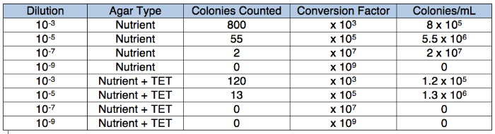
In part one of the experiment in order to quantify and identify organisms the following table summarizes the observations:
http://i35.photobucket.com/albums/d197/corey92/9204068f-5fca-449d-bb11-ba6a23f2e621_zps818c21dc.png
Observations of the agar plates, both TET rich and deficient, had different types of bacteria present. Some colonies were round in shape, others were more differentiated although still circular. Some of the bacterial colonies were yellow in color and others were white with very clear differentiation between. Most of the colonies were raised as opposed to flat and glossy in color. A wet mount of four of the bacterial colonies were observed at 100x magnification before staining; however, photos of the stained bacteria produce a more quality image and representation of the shape, size, and motility of the bacteria and are therefore used in this lab entry. A photo of the colonies can be found below:
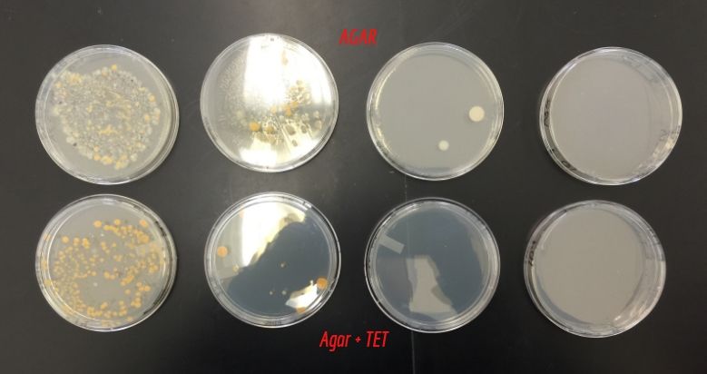
Figure1: All AGAR plates with bacteria from hay infusion. Top Row: AGAR -TET Bottom Row: AGAR +TET
http://i35.photobucket.com/albums/d197/corey92/0445a37a-630a-4297-b18f-d5fbfd44a513_zps6e363789.jpg
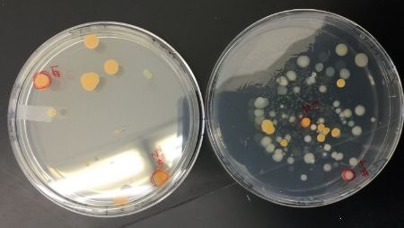
In part 2 of the experiment, an assessment of antibiotic resistance was done via gram staining. Pictures of the gram stain are below:
Figure 3: Image of gram stain slides #1-4
http://i35.photobucket.com/albums/d197/corey92/291c2dee-3e36-4d7d-b0a2-ba09b4f3b728_zps4efd7361.jpg
Figure 4: Slide #1 showing gram positive bacteria sample from TET+ plate (100X)
http://i35.photobucket.com/albums/d197/corey92/60c28e1a-94c9-40d3-ac5d-9f360b9ebb53_zpsd4b74a75.jpg
Figure 5: Slide #2 showing gram positive bacteria sample from TET+ plate (100X)
http://i35.photobucket.com/albums/d197/corey92/65c9e50a-89e2-460b-89a7-88daf4e40af0_zpsfbfd09c8.jpg
Figure 6: Slide #3 showing gram negative bacteria sample from AGAR -TET (100x)
http://i35.photobucket.com/albums/d197/corey92/6d15fba0-5390-47ad-8533-1225bec66ce0_zpse1b58d1c.jpg
A summary of observations of bacteria are as follows:
http://i35.photobucket.com/albums/d197/corey92/ScreenShot2015-02-05at120636AM_zps323b4c0d.png
CONCLUSIONS & FUTURE DIRECTIONS:
Gram positive bacteria are antibiotic resistant based on the observation that only the gram positive bacteria existed on the Agar + TET plates. In terms of the observations made on the plates, observations are in line with the mechanisms of action as discussed in 2001 by Ian Chopra and Marilyn Roberts, "Tetracyclines traverse the outer membrane of gram-negative enteric bacteria through the OmpF and OmpC porin channels, as positively charged cation (probably magnesium)-tetracycline coordination complexes (44, 263). The cationic metal ion-antibiotic complex is attracted by the Donnan potential across the outer membrane, leading to accumulation in the periplasm, where the metal ion-tetracycline complex probably dissociates to liberate uncharged tetracycline, a weakly lipophilic molecule able to diffuse through the lipid bilayer regions of the inner (cytoplasmic) membrane." Also notable was the absence of any organisms classified as archaea in the plates which makes sense because archaea generally thrive in areas of extreme environmental conditions.
- Corey J Salas 22:50, 4 February 2015 (EST):
2.4.15 Excellent lab book entry. Nice detailed description of Hay Infusion and particular protists found within. Logical and well organized. Beautiful diagram of serial dilutions! SK
LAB #2: Identifying Algae and Protists
PURPOSE:
The general purpose of this lab was to practice using a dichotomous key to identify unknown algae and protists from our transect at American University as well as gaining a clearer understanding of the characteristics of algae and protists.
MATERIALS & METHODS:
This lab was divided into three main parts. The first was to assist students in using a dichotomous key, the second was to identify life at the microbiological level in the hay infusion culture prepared the previous week, and third to prepare and plate a serial dilution. In the first part of the lab, students were instructed to go around the room and observe different microscopic slides and use the dichotomous key to attempt to align their observations with the organism deduced using the key. Students were then instructed to prepare a wet mount of known organisms and observe them with the microscope at 4x and 10x magnifications.
In the second part of the lab, samples were taken from the hay infusion culture from the bottom, middle, and top sections arbitrarily assigned by the group members. Wet mounts were prepared and a determination of which protists or algae was undertaken using the dichotomous key. Figures 1-4 below show some of the external, internal and microscopic observations.
The third part of the lab required students to create a serial dilution with a sample of the hay infusion culture and 10mLs of sterile broth. Four test tubes labeled 10^2, 10^4, 10^6, 10^8 with approximately 10mLs of sterile brother were used in the dilution. 100µl of the culture were added to the 10mLs of broth labeled 10^2, 10µl of the broth from the tube labeled 10^2 were then added to the tube labeled 10^6, 100µl of the broth from the tube labeled 10^6 were then added to the tube labeled 10^8 which completed the serial dilution preparation. These dilutions were then plated on the nutrient rich agar plates in two sets, one set with +TET and one set without. See the serial dilution diagram below (Figure 5).
DATA & OBSERVATIONS:
The second portion of the lab yielded the most interesting results as organisms from the group hay infusion culture were being observed. All organisms observed appeared to be motile and were in both the algae and protazoan lineages. None of the organisms were more than 200µl in size and were not visible to the naked eye; therefore an assessment of their loitering near plant matter in the infusion culture is difficult . Observed in the samples placed on the wet mounts were the following:
Upon initial contact with the hay infusion culture, it is notable that the smell had changed from the initial preparation approximately 7 days prior. There was a smell of putrefaction exuding from the jar as well as a white film that had formed in the top portion of the jar - the group assumes this is some type of mold.
http://i35.photobucket.com/albums/d197/corey92/1d54e7bc-d75a-4d97-9a08-8281a6d174a3_zps3e6e7af0.jpg http://i35.photobucket.com/albums/d197/corey92/34c8d0aa-8417-4a12-9c91-9066bac5ee23_zps698186af.jpg
Figure 5: Serial dilution of hay infusion cultures to agar plates.
CONCLUSIONS & FUTURE DIRECTIONS:
Due to the biological nature of our study this semester, it is important to relate what we observe back to what we know about our definition of life. Thinking about the organisms observed in the hay culture, chlamydomonas serve well to bridge the gap between how we define life and how we observe the characteristics of that definition. Chlamydomonas has cells, uses energy (photosynthesis), grow and can reproduce - these are all characteristics that define life as well as characteristics of chlamydomonas itself. Thinking forward about how this culture might grow in the future, depending on environmental conditions, the infusion culture might grow more organisms or could very well kill them off due to lack of resources, it really all depends.
- Corey J Salas 17:16, 28 January 2015 (EST):
1.27.15 Excellent first lab book entry. Well structured and organized. Nice photo. Transect is 20 ft by 20 ft there is a typo in the manual. SK
LAB #1: Biological Life at AU
PURPOSE
The general purpose of the lab observations were to observe biodiversity as it exists in the environment as well as providing a means for a sample in order to create a Hay Infusion Culture which would further allow students to observe biodiversity on a more microbiological level.
MATERIALS & METHODS
During the in class portion of the lab, the volvocine line was observed in order for students to view the evolution of a line as well as to reorient themselves with the use of the microscope.
The materials used in the latter half of the lab were a sterile 50mL conical tube in which a soil/ground vegetation sample was placed as well as a clip board, notebook paper, and pencil in order to take accurate and detailed notes. A cell phone with a camera was also recommended in order to take photographs of the area. Students were randomly assigned a transect and were instructed to then travel to and observe the area, take detailed notes, as well as take a soil sample which was representative of the entire transect. In addition to making notes about the topography and general characteristics of the area biotic and abiotic features of the area were also recorded.
A secondary part of the lab was to prepare a hay infusion culture from the approximately 10-12 grams of the soil/ground vegetation sample mixed with 500mL of deer park brand water and 0.1g of dried milk powder. The mixture was mechanically disrupted with a lid on the jar and allowed to sit for approximately one (1) week. The hay infusion culture will be used to observe, on a microbiological level, the biodiversity present in the assigned transect.
DATA & OBSERVATIONS
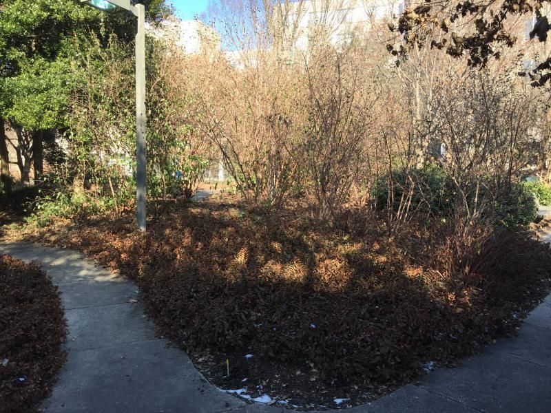
General Transect Characteristics: The transect observed is an approximately 20 ft by 20 ft plot marked with four popsicle sticks. The transect was labeled transect #3 and is not perfectly rectangular in shape, rather one might compare it to the shape of a trapezoid. The ground is relatively mound shaped with consistent elevation in the center of the transect gradually decreasing toward the outer regions of the plot with concrete sidewalks surrounding the edges of the transect. The highest point of the transect is approximately 2.5 feet at its highest point measuring from the level of the concrete sidewalk. The transect is located approximately 20-30 feet from the bender sports arena and is located at approximately 38 degrees N and 77 degrees W. It is located near other areas of the recreational park as well as a major roadway that runs through the American University campus. The area is primarily used as a park area for students, faculty, staff and visitors to American University to relax and perform recreational activities. There are small shrubs, taller trees, and rotting foliage present within the plot as well as a diversity of animals and humans that come through the area at various times.
Biotic Features: 1) Squirrel, 2) Human, 3) Leafy Green Plants 4) Trees, 5) Small bushes
Abiotic Features: 1) Metal light post, 2) plastic meter box, 3) ribbon 4) concrete side walk, 5) stones
CONCLUSIONS & FUTURE DIRECTIONS
Based on the observations of the transect and the sample collected and prepared in the hay infusion culture, it is advantageous to say that there is much biodiversity present in and around the American University campus. It is also important to note that the changing biotic and abiotic features of the transect will be of particular interest to observe as the semester progresses.
--Corey J Salas 22:54, 26 January 2015 (EST)
Test. Wednesday, January 21, 2014. *Corey J Salas 15:02, 21 January 2015 (EST):