User:Dhea Patel/Notebook/Hemoglobin Project/2013/02/05: Difference between revisions

From OpenWetWare
Jump to navigationJump to search
mNo edit summary
Line 57: Line 57:
[[Image:9.8mg_psorbitol_CitrateB_Solid_in_1mL_Ethyl_Acetate_.png]]
[[Image:9.8mg_psorbitol_CitrateB_Solid_in_1mL_Ethyl_Acetate_.png]]
*Fluorescence of "p-sorbitol in Citrate buffer" solid in Ethyl Acetate
[[]]
[[Image:Fluorescence_of_9.8mg_p-sorbitol_in_Citrate_Buffer_Solid_in_1mL_Ethyl_Acetate_.png]]


*UV-vis of "p-sorbitol in Citrate buffer" solid in Water
[[Image:10.0mg_psorbitol_CitrateB_Solid_in_1mL_Water.png]]
[[Image:10.0mg_psorbitol_CitrateB_Solid_in_1mL_Water.png]]
*Fluorescence of "p-sorbitol in Citrate buffer" solid in Water
[[]]
[[Image:Fluorescence_of_10.0mg_p-sorbitol_in_Citrate_Buffer_Solid_in_1mL_Water_.png]]


*UV-vis of "p-sorbitol in Citrate buffer" solid in Chloroform

Revision as of 09:02, 20 February 2013

Project name <html><img src="/images/9/94/Report.png" border="0" /></html> Main project page
<html><img src="/images/c/c3/Resultset_previous.png" border="0" /></html>Previous entry<html>&nbsp;&nbsp;&nbsp;&nbsp;&nbsp;&nbsp;</html>Next entry<html><img src="/images/5/5c/Resultset_next.png" border="0" /></html>

WP:TOC

Objective

  • to finish running samples from Friday
  • to run samples of solid (0.2g p-sorbitol , 0.01 Mb, 0.1g PEG in 10mL citrate buffer) made January 29th.

Description

  1. 10.0mg of the "Myoglobin in Citrate buffer" solid was added to 1mL water
  2. 10.2mg of the "Myoglobin in Citrate buffer" solid was added to 1mL chloroform
  3. 10.0mg of the "p-sorbitol in Citrate buffer" solid was added to 1mL water
  4. 11.4mg of the "p-sorbitol in Citrate buffer" solid was added to 1mL chloroform
    1. the solid remained in a clump and floated to the top of the tube.
  5. 9.8mg of the "p-sorbitol in Citrate buffer" solid was added to 1mL ethyl acetate
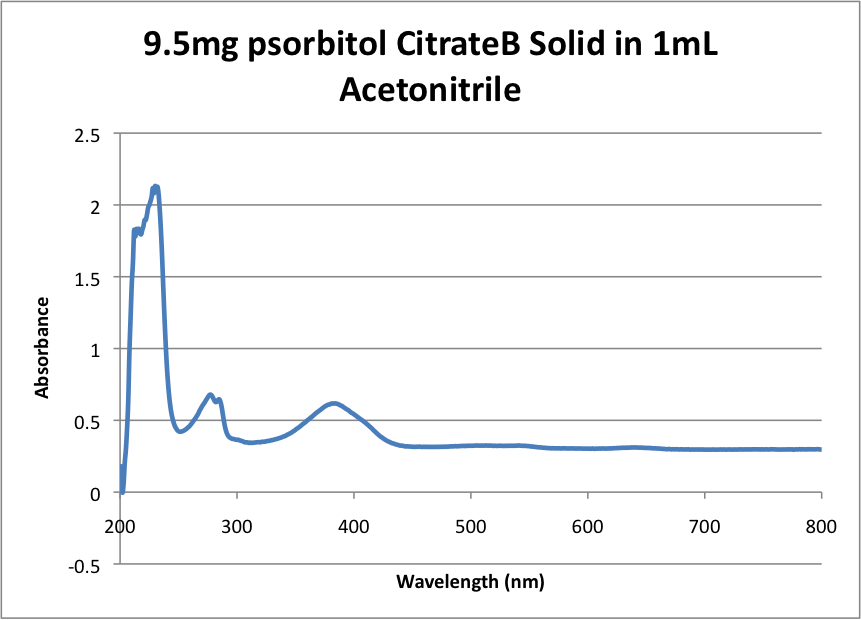
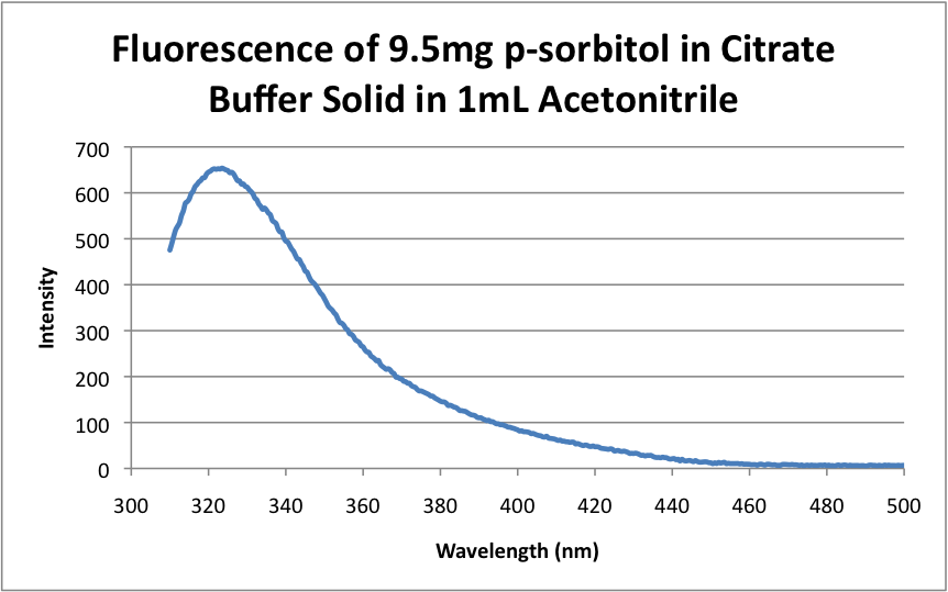
  6. 9.5mg of the "p-sorbitol in Citrate buffer" solid was added to 1mL acetonitrile
  7. 11.9mg of the "p-sorbitol in Citrate buffer" solid was added to 1mL acetone
  8. 9.7mg of the "p-sorbitol in Citrate buffer" solid was added to 1mL methanol
  • all the samples were vortexed and centrifuged before being measured
  • Centrifuge settings were as follows:
    • 13200rpm
    • 0:16 seconds
    • 851 rotor
  • each solvent was used as a blank.
  • The data was corrected for the blank and a corrected baseline.
  • UV-vis scanned from 200-800nm
  • Fluorescence was run with an emission scan of 310-550nm at an excitation of 290nm and scan speed of 100nm/min.

Data

  • Please see last Friday's Data for the UV-vis and Fluorescence spectra of the myoglobin samples.


  • UV-vis of "p-sorbitol in Citrate buffer" solid in MeOH

  • Fluorescence of "p-sorbitol in Citrate buffer" solid in MeOH

[[]]

  • UV-vis of "p-sorbitol in Citrate buffer" solid in Acetone

  • Fluorescence of "p-sorbitol in Citrate buffer" solid in Acetone

[[]]

  • UV-vis of "p-sorbitol in Citrate buffer" solid in Acetonitrile

  • Fluorescence of "p-sorbitol in Citrate buffer" solid in Acetonitrile

  • UV-vis of "p-sorbitol in Citrate buffer" solid in Ethyl Acetate

  • Fluorescence of "p-sorbitol in Citrate buffer" solid in Ethyl Acetate

  • UV-vis of "p-sorbitol in Citrate buffer" solid in Water

  • Fluorescence of "p-sorbitol in Citrate buffer" solid in Water

  • UV-vis of "p-sorbitol in Citrate buffer" solid in Chloroform

  • Fluorescence of "p-sorbitol in Citrate buffer" solid in Chloroform