User:Hayes Braxton Whitney/Notebook/Biology 210 at AU: Difference between revisions
No edit summary |
No edit summary |
||
| (2 intermediate revisions by the same user not shown) | |||
| Line 9: | Line 9: | ||
[[Image:table1zebra.jpg]] | [[Image:table1zebra.jpg]] | ||
After gaining knowledge on embryonic development, the scientists set up the Zebrafish experiment. 20 Zebrafish embryos were placed into a control dish consisting of 20mL of normal Deer Park water. Another 20 embryos were separately placed in a dish consisting of 20mL of normal water and 4mL of nicotine was then added. The scientists hypothesized that the Zebrafish exposed to nicotine would have a higher energy level while embryonic and after hatching. Observations of the Zebrafish will be made over the next two weeks. Observations will be added upon conclusion of the experiment. Day 2 and day 5 of the experiment have been observation days, day 5 consisted of cleaning and feeding the Zebrafish, results from those observations are shown in the table below. | After gaining knowledge on embryonic development, the scientists set up the Zebrafish experiment. 20 Zebrafish embryos were placed into a control dish consisting of 20mL of normal Deer Park water. Another 20 embryos were separately placed in a dish consisting of 20mL of normal water and 4mL of nicotine was then added. The scientists hypothesized that the Zebrafish exposed to nicotine would have a higher energy level while embryonic and after hatching. Observations of the Zebrafish will be made over the next two weeks. Observations will be added upon conclusion of the experiment. Day 2 and day 5 of the experiment have been observation days, day 5 consisted of cleaning and feeding the Zebrafish, results from those observations are shown in the table below. | ||
[[Image:Exzebra.jpg]] | [[Image:Exzebra.jpg]] | ||
[[Image:table1213123.jpg]] | |||
| Line 41: | Line 42: | ||
ACGANNCAACCCCTGTNNTTAGTTN | ACGANNCAACCCCTGTNNTTAGTTN | ||
[[Image:blast2.jpg]] | |||
Sequence File : MB2-For_16S.seq | Sequence File : MB2-For_16S.seq | ||
| Line 61: | Line 62: | ||
GCAACNAGCNCAACCCCTGNCACTANTNGCNNN | GCAACNAGCNCAACCCCTGNCACTANTNGCNNN | ||
[[Image:blast1.jpg]] | |||
Feb 11th, 2015 | Feb 11th, 2015 | ||
Latest revision as of 11:23, 26 February 2015
Feb 18th 2015 Lab 6: Embryology & Zebrafish Development
The purpose of Lab 6 was to further understand the stages of embryonic development through observation of Zebrafish. Also, the scientists set up an experiment to begin to understand how the environmental conditions will affect the development of the Zebrafish.
Procedures had the scientist observe the embryonic development of a starfish embryo, a frog embryo, and a chick embryo respectively. Table 1 shows the comparisons of the three observed embryos, the fourth observation being the embryonic development of a human.
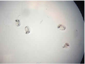
After gaining knowledge on embryonic development, the scientists set up the Zebrafish experiment. 20 Zebrafish embryos were placed into a control dish consisting of 20mL of normal Deer Park water. Another 20 embryos were separately placed in a dish consisting of 20mL of normal water and 4mL of nicotine was then added. The scientists hypothesized that the Zebrafish exposed to nicotine would have a higher energy level while embryonic and after hatching. Observations of the Zebrafish will be made over the next two weeks. Observations will be added upon conclusion of the experiment. Day 2 and day 5 of the experiment have been observation days, day 5 consisted of cleaning and feeding the Zebrafish, results from those observations are shown in the table below.
Feb. 25th, 2015
Blast
Sequence File : MB1-For_16S.seq
>MB1-For_16S_A01.ab1 NNNNNNNNNNNNNNANNNGCAGTCGGANNGGNNGNNNNNNNNNNNNNCGGCNGAGACCGGCGCACGGGTGCGTAACGCGT ATGCAATCTACCTTTTACAGAGGGATAGCCCAGAGAAATTTGGATTAATACCTCATAGTATAACACAATCGCATGATTGA GTTATTAAAGTCACAACGGTAAAAGATGAGCATGCGTCCCATTAGCTAGTTGGTAAGGTAACGGCTTACCAAGGCTACGA TGGGTAGGGGTCCTGAGAGGGAGATCCCCCACACTGGTACTGAGACACGGACCAGACTCCTACGGGAGGCAGCAGTGAGG AATATTGGACAATGGGCGCAAGCCTGATCCAGCCATGCCGCGTGCAGGATGACGGTCCTATGGATTGTAAACTGCTTTTG TACGAGAAGAAACACCTCTTCGTGTAGGGACTTGACGGTATCGTAAGAATAAGGATCGGCTAACTCCGTGCCAGCAGCCG CGGTAATACGGAGGATCCAAGCGTTATCCGGAATCATTGGGTTTAAAGGGTCCGTAGGCGGTTTAGTAAGTCAGTGGTGA AAGCCCATCGCTCAACGGTGGAACGGCCATTGATACTGCTGAACTTGAATTATTAGGAAGTAACTAGAATATGTAGTGTA GCGGTGAAATGCTTAGAGATTACATGGAATACCAATTGCGAAGGCAGGTTACTACTAATGGATTGACGCTGATGGACGAA AGCGTGGGTAGCGAACAGGATTAGATACCCTGGTAGTCCACGCCGTAACGATGGATACTAGCTGTTGGGAGCAATTTCAG TGGCTAAGCGAAAGTGATAAGTATCCCACCTGGGGAGTACGTTCGCAAGAATGAAACTCNNGGAATTGACGGGGGCCCGC ACAAGCGGTGGAGCATGTGGTTTAATTCNATGATACNCGAGGAACCTTACCAANGCTTAAATGTANTGTGNNCCGATNTG GANCAGATCTTTCGCANACAAATTACAANNGCTGCATGGTNGTCNTCAGCTCGTGCCGTGAGNNNCNGNTAANTCCNATA ACGANNCAACCCCTGTNNTTAGTTN
Sequence File : MB2-For_16S.seq
>MB2-For_16S_B01.ab1 NNNNNNNNNNNGNNNANNCATGCAAGCCGAGCGGTAGAGANCTTTCGGGATCTTGAGAGCGGCGTACGGGTGCGGAACAC GTGTGCAACCTGCCTTTATCAGGGGGATAGCCTTTCGAAAGGAAGATTAATACCCCATAATATATTGAATGGCATCATTT GATATTGAAAACTCCGGTGGATAGAGATGGGCACGCGCAAGATTAGATAGTTGGTAGGGTAACGGCCTACCAAGTCAGTG ATCTTTAGGGGGCCTGAGAGGGTGATCCCCCACACTGGTACTGAGACACGGACCAGACTCCTACGGGAGGCAGCAGTGAG GAATATTGGACAATGGGTGAGAGCCTGATCCAGCCATCCCGCGTGAAGGACGACGGCCCTATGGGTTGTAAACTTCTTTT GTATAGGGATAAACCTTTCCACGTGTGGAAAGCTGAAGGTACTATACGAATAAGCACCGGCTAACTCCGTGCCAGCAGCC GCGGTAATACGGAGGGTGCAAGCGTTATCCGGATTTATTGGGTTTAAAGGGTCCGTAGGCGGATCTGTAAGTCAGTGGTG AAATCTCATAGCTTAACTATGAAACTGCCATTGATACTGCAGGTCTTGAGTAAAGTAGAAGTGGCTGGAATAAGTAGTGT AGCGGTGAAATGCATAGATATTACTTANAACACCAATTGCGAAGGCAGGTCACTATGTTTNANNNNACGCTNATAGGACG AAAGCGTGGGGAGCGAACAGGATTAGATACCCTGGTAGTCCACGCCGTAAACGATGCTAACTCGTTTTTGGGTCTTCGGA TTCAGAGACTAAGCGAAAGTGATAAGTTAGCCACCTGGGGAGTACGTTCGCAAGAATGAAACTCAAAGGAATTGACGGGG GCCCGCACAAGCGGTGGATTATGTGGTTTAATTCGATGATACGCGAGGAACCTTACCAANNCTTAAATGGGAATTGACAG GTTTANAAATANNCTTTTCTTCGNANATTTTCNNNGCTGCATGGNNTCGTCAGCTCNTGCCNTGAGTGTTNGNNAGTCCT GCAACNAGCNCAACCCCTGNCACTANTNGCNNN
Feb 11th, 2015
Lab 5: Invertebrates
Lab 5 was designed for the scientists to begin to understand the importance of invertebrates and to learn how evolution of simple systems into complex ones takes place.
Procedure 1 had the scientists observing the Acoelomates, Pseudocoelomates, and Coelomates. The movements were observed and documented as follows: The acoelomates moved in a snake-like motion, and were shaped in a long, lean manner. The Pseudocoelomates were shaped in a more round fashion, and moved spirally. The Coelomates moved more fluidly than the others, in a smooth fashion, and shaped similarly to the Coelomates.
Procedure 2 had the scientists observing the arthropod examples to make it easier to identify organisms from the Burlese funnel transect sample. Procedure three had the scientists take two samples from the Burlese funnel and placed into two separate observation dishes. There was an abundance of invertebrate life in the samples, as shown in the pictures below. Five invertebrates were identified as follows, 1. Soil Mite 2. Ground Spider 3. Spring-Tail 4. Soil Mite 5. Proturan. The sizes of the specimens varied, the smallest being the Soil mites at .4mm and the largest being the ground spider at 10mm observed. The specific descriptions are shown in the table below.
Feb. 4th, 2015 Lab 4: Plantae and Fungi
Lab 4 was designed for the scientists to understand the function of plants and the diversity they offer to the surrounding ecosystem, and to further understand the functionality of Fungi and it’s interaction within Transect 1. The first procedure consisted of the scientists collecting two separate samples from Transect 1. The first sample collected was 500g of leaves and assorted fauna from the transect and placed in a large Ziploc bag for transport. The second Ziploc bag was carefully filled with five separate samples of present plant species within Transect 1. These five samples are shown below in respect to the quadrant location within the transect. The 500g Ziploc bag containing the leave samples was subsequently emptied into a Berlese funnel for invertebrate collection, which will be completed in Lab 5.
The five plants collected from the transect quadrants are shown below, and are numbered in respect to their transect location (1-5). Plant 1 is commonly noted as a Cattail, and is one of the most prevalent plants within the transect. The stalk is tall and thin, approximately four-six feet in height depending on the season and damage done to the plant. Since it is winter, the plants are not thriving in this weather. The head of the cattail is quite delicate and when irritated lets out multiple white flower buds. This plant falls into the typha genus. Plant two is simply moss, it was located close to the sidewalk and is moist with mud/dirt sediment. This plant would fall into the bryophyte genus. Plant three was actually labeled by the University as a “red cardinal flower bush”, indicated by the red berry growth on the plant’s long, narrow stems. This plant is part of the lobelia genus. Plant four is large, similar to plant one. It has long, narrow stems and the flowers on the end of the stems have seedlings in pods. This plant is could be identified as an angiosperm because of the budding flowers. Plant five, also a angiosperm, has smaller budding seedlings on the end of the branches, similar to plant four.
In procedure two, the scientists compared the lily (angiosperm) and the moss (Mnium). The moss grows on the ground, and does not grow vertically like the lily. The plants were ordered from tallest to shortest, plant one (cattail) being the tallest. Cross sections were viewed and the cattail revealed a narrow, hollow stem that would break easily under pressure. Due to seasonal weather, the scientists had difficulty making observations of non-bush plants. Plant two was the only other easily viewable plant because of its intense features, and was similar to the moss viewed earlier in comparison with the lily. Below is the Transect Sample Plant observations.
In the last procedure, the scientists set up the Berlese funnel was set up for invertebrate extraction. In order to do this, a conical tube was securely attached to the bottom of the Berlese funnel after it was filled with 25mL of ethanol. The funnel was secured to the stand and 500g of leaves were placed into the funnel. Once complete, the funnel was placed under light and covered with aluminum foil to maximize heat input. The invertebrates collected from the 500g sample will be observed in lab 5.
1/28/2015 Lab 3: Microbiology and Identifying Bacteria with DNA Sequences
The objectives of lab 3 were to understand how bacteria works in certain environments, observe the bacteria’s resistance to antibiotics, and using PCR to identify species of bacteria through viewing DNA. Transect 1’s simple, stable environment is not one that Archaea can thrive in; they require more extreme temperatures that come with harsh environments unlike the local transects.
Purpose
Before beginning procedure 1, an observation of transect 1’s Hay Infusion Culture was made. The Hay Infusion’s smell has gotten much more distinct and putrid over the last week. There has also been a slight thickening of the culture on the top layer, and water appears to have evaporated over time. This observation is in line with the previous hypothesis that allowing the Hay Infusion Culture to sit for a week in the lab environment would cause it to develop further and achieve the smell and visual appearance mentioned above. If allowed to continue, the Hay Infusion Culture would continue to develop more putrid smells until the water was completely evaporated.
Materials & Methods The agar plates that were inoculated from the Hay Infusion cultures were observed in Lab 3 to determine how much growth, if any, had occurred. Each agar plate was observed and colonies were specifically counted and recorded as shown in Table 1 below. In observing plates without antibiotics versus pates with antibiotics, it was visible that although all of the plates had growth, the ones with the antibacterial tetracycline had far less bacterial growth in comparison to fungal growth. This is indicative of the bacteria growing far more resistant to antibiotics. The observations indicate 3-4 species of bacteria unaffected by the tetracycline.
Data & Observations/ Conclusion
Procedure 3 involved viewing bacterial cell morphology, first at 40x then after becoming comfortable with procedure, the scientists proceeded to use the 100x oil immersion objective lens. Four samples of microorganisms were then prepared and viewed; two from the nutrient agar plates and two from the tetracycline plates. While under 100x oil immersion, the 10-7 colony showed no movement and had light colorations of orange and blue approx. 1 cm in diameter. The 10-9 nutrient agar plates showed a colony with no movement and purple coloration approx. 2 cm in diameter. The 10-7 tetracycline plates showed a raised, white circular smooth pattern approx. 3 cm in diameter. The 10-9 tetracycline plate showed a light orange, circulate smooth raised sample approx., 2cm in diameter, surrounded by fungus. All of this information is shown in Table 2 below.
Gram stains were then created for observation. A bacteria sample was scraped from the agar and a drop of water was added on a slide. The slides were labeled accordingly, 10-7 nut., 10-9 nut., 10-7 tet. and 10-9 tet. The bacterial slide was passed through the flame of the Bunsen burner three times to heat fix the slide. The smear was then covered with crystal for one minute’s time and then rinsed. Gram’s iodine mordant was then applied to the smear and let sit for one minute. The stain was then washed off. The color was then removed by immersing the smear with 95% alcohol for approx. 20 seconds. The smear was covered with safranin stain for 20 seconds and rinsed. After the completion of the gram stain, the sample is viewed under 40x and 100x, respectively.
Procedure 4 saw the setup for PCR 16S sequencing. A single colony of bacteria was placed in 100ul of water in a sterile tube, which is then incubated to 100 degrees C for ten minutes. After completing this, the samples were transferred to a centrifuge for five minutes at 13,400rpm. 20ul of primer/water mixture was added to a PCR tube and mixed. After centrifugation, 5ul of supernatant was taken from the sample and added to the 16S PCR reaction. This is setting up next week’s lab where we will run the PCR products on an agarose gel, where we will be able to identify our bacteria.
January 22nd, 2015
Lab 2: Identifying Algae & Protists
Great description on the protists, the images made what you were looking at very clear. Make sure you have observations on your hay infusion so that you can easily remember things when you are putting all the information together. You have a good start to the notebook, but are still missing a couple things. Keep up the good separation of the information though. ML
Purpose The purpose of Lab two was to further understand the usage of the dichotomous key and being able to implement it as a tool to make conclusions on observed Algae and Protists from the group transect.
Materials and Methods The first part of Lab 2 was learning how to use the dichotomous key to correctly identify organisms taken from transect 1. The second part of the lab involved taking samples from transects 1’s hay infusion and creating wet mount slides for viewing through the microscope. The sample was given a week to ferment. The wet mounts were viewed and observations documented. Lastly, 100-microliter sample dilutions were taken from our hay infusion culture from transect 1 and placed into 100mL tubes of sterile broth. This was done four times from the initial tube, creating three additional tubes of subsequently more dilution. After this, agar plates were filled with 100mL from each tube was transferred to each plate-making six plates total. The sample was evenly spread around and placed aside for further observation.
Data and Observations
Six organisms were observed and documented in the table shown below. In procedure two, a sample was taken from transect 1’s hay infusion and organisms were viewed to track the organismal growth. In the third procedure, agar plates were created for viewing during week 3 lab.
Conclusions and Future Directions In conclusion, the fermentation of transect 1’s hay infusion was viewed and showed organismal growth throughout. Future experiments will involve viewing the more in depth growth over the next four weeks of fermentation.
January 14th, 2015
Lab 1: Biological Life at American University
This information is very well put together and you should continue to record data at this level, but there is no information about your transect in your notebook other than the picture at the end. Make sure you follow the directions in the red boxes when writing your notebook. ML
The purpose of Lab 1 was to understand how natural selection plays a role in the group transect, as well as observe procured samples from our transect under microscope.
Materials and Methods The first part of Lab 1 involved viewing of the Volvocine Line under microscope. The cells viewed were Volvox, Gonium, and Chlamydomonas, shown below (Image1). The number of cells varies throughout the Volovcine Line, but typically Chlamydomonas shows a single cell, Gonium presents from the 4-32 cell range, and Volvox appears as a great ball of cells. The results shown below indicated this in subdued form. After the Volvocine Line cells were viewed, the second part of the experiment involved moving to the assigned groups transect. A 50ml transect sample was taken and the procured samples documented (Image 2). Upon returning to the lab, a Hay Infusion Culture was created using 10 grams of the transect sample and 500mLs of Deer Park water. After that, 0.1g of powdered milk was added to the jar for nutrients for the living organisms and was mixed to evenly distribute the powdered milk throughout the sample.
Data and Observations Images 1 and 2 show the data and observations from Lab 1. Image 3 shows the Scientist in front of the transect for measurement purposes.
Conclusions and Future Directions Observations will continue to be made of the transect sample over the coming weeks. It is likely that we will see vast organismal growth.
HBW GRAVEL PIT


