User:Jessica Kerpez/Notebook/Biology 210 at AU: Difference between revisions
No edit summary |
No edit summary |
||
| Line 7: | Line 7: | ||
The objectives of this lab were to learn and compare embryonic development in an array of organisms and set up an experiment to study how environmental conditions affect embryonic development. Also, zebrafish can be used as a model for comparison to larger vertebrae development. For this specific experiment, zebra fish were used to observe the stages of embryonic development because they are clear and easy to see and because they share similar embryonic development to humans. In the experiment, one group of zebrafish was the control group in normal deerpark water and one group of zebrafish were treated with 25 mg of caffeine. The hypothesis was that zebrafish treated with caffeine will display defects in their development, resulting in spastic motor movement and mutations in the eyes and body length. | The objectives of this lab were to learn and compare embryonic development in an array of organisms and set up an experiment to study how environmental conditions affect embryonic development. Also, zebrafish can be used as a model for comparison to larger vertebrae development. For this specific experiment, zebra fish were used to observe the stages of embryonic development because they are clear and easy to see and because they share similar embryonic development to humans. In the experiment, one group of zebrafish was the control group in normal deerpark water and one group of zebrafish were treated with 25 mg of caffeine. The hypothesis was that zebrafish treated with caffeine will display defects in their development, resulting in spastic motor movement and mutations in the eyes and body length. | ||
[[State the Specific Steps]] | |||
1. Twenty zebrafish embryos were placed in 20 mLs deerpark water and twenty zebrafish embryos were placed in 20 mLs of 25 mg of caffeine per liter of water solution. | 1. Twenty zebrafish embryos were placed in 20 mLs deerpark water and twenty zebrafish embryos were placed in 20 mLs of 25 mg of caffeine per liter of water solution. | ||
| Line 17: | Line 17: | ||
4. On day 14, final data was collected on the organisms including how many were still alive and any major the differences between the treated and control group. 1 drop of triacine solution was added per mL of water, and the TA added paraformaldehyde. | 4. On day 14, final data was collected on the organisms including how many were still alive and any major the differences between the treated and control group. 1 drop of triacine solution was added per mL of water, and the TA added paraformaldehyde. | ||
[[Include Raw Data]] | |||
Data is listed below. | Data is listed below. | ||
[[State Conclusions and Future Plans]] | |||
Consistent with the hypothesis created about the zebrafish, the group treated with caffeine demonstrated a significantly lower rate of survival than the embryos placed in solely deer-park water. 12 were dead and 8 were alive from the caffeine group, compared to 5 dead and 15 alive from the group placed in only water. Also, the fish swam with erratic movement, the reaction time to stimulus was severely off in the group were caffeine was present in the water. The distance of the eyes was significantly larger in the caffeine treated group, and the body length was somewhat shorter in the treated group. In eye distance in the control group was 15 um apart, and the distance of eye length for the treated group was significantly larger at 25 um. The body length of the control group was 412 um, while the treated group was 380 um on average. | Consistent with the hypothesis created about the zebrafish, the group treated with caffeine demonstrated a significantly lower rate of survival than the embryos placed in solely deer-park water. 12 were dead and 8 were alive from the caffeine group, compared to 5 dead and 15 alive from the group placed in only water. Also, the fish swam with erratic movement, the reaction time to stimulus was severely off in the group were caffeine was present in the water. The distance of the eyes was significantly larger in the caffeine treated group, and the body length was somewhat shorter in the treated group. In eye distance in the control group was 15 um apart, and the distance of eye length for the treated group was significantly larger at 25 um. The body length of the control group was 412 um, while the treated group was 380 um on average. | ||
[[Image:Picture1biotablex.png]] | |||
[[Image:Picture2biotab.png]] | |||
'''February 24, 2013''' | '''February 24, 2013''' | ||
'''J.K.''' | |||
LAB 5: Invertebrates | LAB 5: Invertebrates | ||
Latest revision as of 11:28, 25 April 2014
April 24, 2014
Lab 6: Embryology & Zebrafish Development
State the question/problem/objective
The objectives of this lab were to learn and compare embryonic development in an array of organisms and set up an experiment to study how environmental conditions affect embryonic development. Also, zebrafish can be used as a model for comparison to larger vertebrae development. For this specific experiment, zebra fish were used to observe the stages of embryonic development because they are clear and easy to see and because they share similar embryonic development to humans. In the experiment, one group of zebrafish was the control group in normal deerpark water and one group of zebrafish were treated with 25 mg of caffeine. The hypothesis was that zebrafish treated with caffeine will display defects in their development, resulting in spastic motor movement and mutations in the eyes and body length.
1. Twenty zebrafish embryos were placed in 20 mLs deerpark water and twenty zebrafish embryos were placed in 20 mLs of 25 mg of caffeine per liter of water solution.
2.These embryos were observed four times over the course of two weeks, and observations were taken of how many were still alive, what specific features had developed over time, and any other features.
3.On day 7, three embryos were to be fixated in the water so that they could be observed the following week.
4. On day 14, final data was collected on the organisms including how many were still alive and any major the differences between the treated and control group. 1 drop of triacine solution was added per mL of water, and the TA added paraformaldehyde.
Data is listed below.
State Conclusions and Future Plans
Consistent with the hypothesis created about the zebrafish, the group treated with caffeine demonstrated a significantly lower rate of survival than the embryos placed in solely deer-park water. 12 were dead and 8 were alive from the caffeine group, compared to 5 dead and 15 alive from the group placed in only water. Also, the fish swam with erratic movement, the reaction time to stimulus was severely off in the group were caffeine was present in the water. The distance of the eyes was significantly larger in the caffeine treated group, and the body length was somewhat shorter in the treated group. In eye distance in the control group was 15 um apart, and the distance of eye length for the treated group was significantly larger at 25 um. The body length of the control group was 412 um, while the treated group was 380 um on average.
February 24, 2013
J.K.
LAB 5: Invertebrates
State the question/problem/objective
The objectives of this lab were to understand the importance of inverterbrates, and to learn how simple systems were able to evolve into more complex systems. By observing acoelomates, pseudocoelmoates, and coelomates, the way that body structure and movement correlate between species could be appreciated. Then, we studied soil invertebrates from our transect to better understand invertebrates. I hypothesized that by observing the soil invertebrates from our transect, the diversity and importance of invertebrates could be appreciated. Soil animals help in breaking down and recycling decomposing plant and animal material.
1.The Acoelomates, Pseudocoelmoates, and Coelomates were observed under the microscope and their movement and body structure was evaluated.
2.The Berlese setup was broken down and the preservative solution was transferred to a Petri dish in order to be examined under the dissecting microscope.
3.The invertebrates from the Berlese Funnel were identified using dichotomous keys.
4.We were able to find a total of three organisms: the ground spider, millipede, and centipede. The length and description of these organisms were recorded.
5. Verterbrates who could inhabit or pass through the transect were considered. Characteristics of the five vertebrates (two being bird species) were identified such as the classification and the abiotic/biotic elements. A food web was created based upon the observed organisms.
Data is shown below.
State conclusions and future plans
The objectives of this lab were explicitly addressed in the procedure. In our Berlese Funnel, we were able to identify a total of three organisms: the ground spider, millipede, and centipede. The Acoelomates, Pseudocoelmoates, and Coelomates were observed under the microscope and their movement and body structure was evaluated. Most soil inverterbrates are anthropods. I was surprised that we were able to identify larger arthropods that live on the soil surface and no micro-arthropods. Larger arthropods, like the millipedes, centipedes, and spiders we were able to identify live on the soil surface. Soil animals are predators while others can suck fluid from roots, consume fungi, or feast on decaying plant material. All of them help in the process of breaking down and recycling decomposing plant and animal matter.
Acoelomates move by gliding, and lack a coelom. Therefore, they are lacking support. Acoelomates have no body cavity at all. They are not as complex in their muscle movement. Pseudocoelomates move by shifting next to each other, in a less defined way than the coelomates. They are not as complex in their muscle movement than coelomates, but they appear somewhat more complex than the acoelomates. The coelomates appear to have a more complex movement, and move by a series of muscle contractions that move their bodies. Coelomates have a fluid filled body cavity called the coelom that has a mesoderm lining. Vertebrates are coelomates.
Table of Organisms:
1. Ground Spider LL- 1 mm in length. The ground spider has no wings, no antennae, 2 body segments, and 8 legs.
2. Millipede- 4.5 mm in length. The millipede has no wings, 1 pair of antennae, several body segments, 2 pairs of legs on each side, and a round body.
3. Centipede- 5 mm in length. The centipede has no wings, 1 pair of antennae, several body segments, 1 pair of legs on each side, and a flat body.
- We were not able to find any more organisms.
The size range of the organisms we measured was from 1 mm in length to 5 mm in length, a 4 mm difference. The largest organism we discovered was the centipede, and the smallest organism was the ground spider. The organisms that were most common was the centipede.
Some vertebrates that might inhabit the transect:
Crows (Chordata, Aves, Passeriformes, Corvidae, Corvus, Corvus brachyrhynchos)
Biotic Advantage: Invertebrates for food, such as worms Abiotic Advantage: Man made feeder
Cardinals (Chordata, Aves, Passeriformes, Passeri, Cardinlidae, Periporphyrus, Cardinalis)
Biotic Advantage: Invertebrates for food, such as worms Abiotic Advantage: Man made feeder, soil
Squirrels (Chordata, Mammalia, Rodentia, Sciuridae, Sciurus, Sciurus carolinensis)
Biotic Advantage: Nuts, seeds, green plants for food Abiotic Advantage: Water feeder, soil
Rabbits (Chordata, Mammalia, Lagomorpha, Leporidae, Oryctolagus, Cuniculus)
Biotic Advantage: Grass or plants for food Abiotic Advantage: Water feeder, soil
Common Frog (Chordata, Amphibia, Anura, Ranidae, Rana, Rana)
Biotic Advantage: Presence of bugs, worms Abiotic Advantage: soil
J.K
February 20, 2013
LAB 4: Plantae and Fungi
State the question/problem/objectives
The objective of this lab was to understand the characteristics and diversity of plants, and to appreciate the function and importance of Fungi. In order to fulfill these objectives, five plants were collected from our transect and evaluated in terms of vascularization, leaves and special characteristics, and reproductive elements. Also, slides were observed and types of Fungi were identified, such as the Ascomycota, which was possible to be identified because of the ascus spores that were visible under the microscope. I hypothesized that because of the vast diversity that exists in plants, all of the matter collected in the transect would display differences in terms of vascularization, special characteristics, and reproductive elements. If the plants were monocot and dicot, displayed different reproductive processes, and different lengths and phenotypes, then diversity will be displayed among the transect.
1.3 zip-lock bags and were obtained, and then we proceeded to the transect.
2.A leaf little sample was obtained from the transect from an area in the middle with softer soil and a variety of dead leaves. About 500g of litter was placed into one bag to be used for the Berlese funnel for next lab.
3.Five representative samples were collected from the transect: a pine-cone, a black cotton-wood, red maple, american elm tree, and grass.
4.The characteristics of a Bryophyte moss, Mnium, and the angiosperm, Lilum, were observed under the microscope. The five samples from the transect were also evaluated under the microscope. All of the seven samples were examined in terms of vascularization, specialized structures, and reproduction.
5.The five plants were described in Table 1. We also determined whether or not the seeds were monocot or dicot.
6.Samples of various fungi were also observed under the microscope and determined to be Zygomycota, Ascomycota, or Basidiomycota.
7.To prepare the Berlese Funnel to collect invertebrates for next lab, 25 mL of the 50:50 ethanol/water solution was poured into the flask.
8.A piece of screening material was placed into the bottom of the funnel, and the sides were tapes so that the leaf litter could not fall into the preservative.
9.The funnel was placed into the neck of the square-sided bottle.
10.The leaf litter sample was put into the top of the funnel, and the entire Berlese Funnel was covered in foil. A 40 watt lamp was set in place above the funnel with an incandescent bulb a few inches from the top of the leaf litter.
11.The lighted set up and Berlese funnel was set up on the lab bench for a week.
Raw data is shown below.
State conclusions and Future Plans
The pinecone, black cotton-wood, red maple, american elm tree, and grass all displayed different shapes and sizes as well as reproductive parts/seeds as was predicted. In this way, the experiment was explicit in addressing the objectives. However, one thing I found somewhat surprising was that most the samples were dicot in terms of vascularization. Monocots have one seed leaf while dicots contain two seed leaves. The samples were identified as dicots due to two cotyledon, the broad leaf or network of veins, and vascular bundles in a ring observed under the compound microscope. Using the microscope, we were also able to learn more about fungi by observing species that were Zygomycota, Ascomycota, and Basidiomycota.
Fungi sporangi are a cell that contains spores, sporangium can be the site of meiosis or mitosis. They are significant because the sporangium must enclose its spores until they are mature and ready to disperse. Asexual sporangia are commonly made by the Chytridomycota and Zygomycota.
I believe this is a fungus because of the spores that open up, the "ascus" spores.
J.K.
"February 20, 2013"
LAB 3: Microbiology and Identifying Bacteria with DNA
State the question/problem/objective
The objectives of this lab was to understand the characteristics of bacteria, to observe antibiotic resistance, and to understand how DNA sequences are used to identify species. The prokaryotes grouped in the domain bacteria were studied, consisting of the Proteobacteria, Chlamydiae, Spirochetes, Actinobacteria, Firmicutres, and the photosynthesizing Cyanobacteria. To examine bacteria, the colony morphology, motility, shape, and subcategories were evaluated. In order to identify bacteria, the gram stain was used. Gram positive looks purple while Gram negative looks red. An important part of this lab was amplifying the 16S gene. Tetracycline inhibits translation by binding to the 16S sub-unit of the 30S prokaryotic ribosomal sub-unit. I hypothesize that the plate with tetracycline with have a reddish color and there will be no purple on the plate. I predict that the plate with tetracycline will have a smaller amount of bacteria because the tetracycline makes it difficult for translation to occur.
1.Lab began by preparing a naive wet mount and a gram stain of two well defined colonies from the nutrient agar plate and two from the tetracycline plate. The four colonies were labeled properly.
2.Prepared slides containing samples of bacillus, coccus, and spirillum shaped bacteria were observed for practice using the 40x and 100x oil immersion objective lens.
3.Four samples of well-isolated microorganisms were selected; two from the nutrient agar plate and two from the nutrient agar plus tetracycline plate.
4.To make a wet mount for each of the samples, a loop was sterilized over a flame and used to scrape up a tiny amount of growth from the surface of the agar, and mixed with a small drop of water. A coverslip was placed over the drop.
5.A second drop was placed on a second slide and smudged with a small amount of the colony in the drop over the surface in order to dry. The area underneath this slide was identified with a red wax pencil in order to be used for the gram stain.
6.The wet mounts were observed under the 10x and the 40x objective. The cell shapes and motility of the organisms were determined.
7.The four group preps were gram stained after they were properly labeled. The slide was passed through a flame three times with the bacterial smear side up in order to dry the slide.
8.A drop of crystal violet covered the smear for one minute, and then the stain was rinsed off using water.
9.Gram's iodine mordant covered the smear for one minute and was gently rinsed.
10.The smear was flooded with 95% alcohol for 10-20 seconds and rinsed in order for decolorization to occur.
11.The smear was covered with safranin stain for 20-30 seconds and rinsed gently.
12. Any excess water was dried with a paper towel.
13.The samples were observed at low magnification and observed under the 40x objective. All observations of cell morphology were recorded.
14.One plate with tetracycline and one without were selected and the 16S rRNA gene was isolated.
15.For the PCR protocal, a small amount of the colonies were placed into their own sterile tube of 100 uL and heated in the heat block for 10 minutes.
16.The solution was spun for 1 minute at 13.5 K RPM.
17.20 uL of the supermix (primers) were added to the PCR tube and 2 uL of the DNA was added to a PCR tube. Then, the solution was placed into the thermocycler.
Data shown below
State Conclusions and Future Plans
On average, the net effect of tetracycline on the total number of the bacteria and colony was much smaller compared to the nutrient agar plate. This occurred because tetracycline inhibited enzyme reactions required for bacterial cells to begin translation. The plate without tetracycline appeared purple, and a mix of oranges was seen on the plate with tetracycline. These results were logical and consistent with what was expected because purple is not resistant to the antibiotic, but yellow, orange, and white are. Also, it was harder for bacteria to survive when tetracycline was present because translation becomes difficult to occur. The characteristics of antibiotic resistance were explicitly witnessed during this procedure.
Yes, the Archaea species will have grown onto the agar plates. Since they are extremophiles, they can grow in a variety of unique environments, including areas in the ocean and on the mainland. For instance, plankton reside in a lot of aquatic environments.
The appearance or smell of the Hay Infusion Culture would change week to week because of increased life forms, such as fungi, bacteria, or other substances. For instance, in our Hay Infusion Culture there was an increase in the amount of filament on the top layer of the water and a deeper more potent stench.
Yes, I see differences in the colony types between the plates with and without antibiotics. There was no purple on the plate without tetracycline, and a mix of oranges on the plate with tetracycline. This occurred because purple is not resistant to the antibiotic, but yellow, orange, and white are. This indicates that the plate with tetracycline had bacteria that could not be resistant. The net effect of tetracycline on the total number or the bacteria and fungi was that there was a smaller amount. It is harder for the bacteria to survive in this environment, because the tetracycline makes it difficult for translation to occur. There are 3 species unaffected by tetracycline: orange, yellow, and white.
Tetracycline inhibit enzyme reactions needed for bacterial cells. It works by binding to 30S ribosome of bacteria, and then stops the attachment of the aminoacyl tRNA to the RNA-ribosome complex. The types of bacteria that are sensitive to this antibiotic are E. coli, Haemophilus influenzae, Mycobacterium tuberculosis, and Pseudomonas aeruginosa.
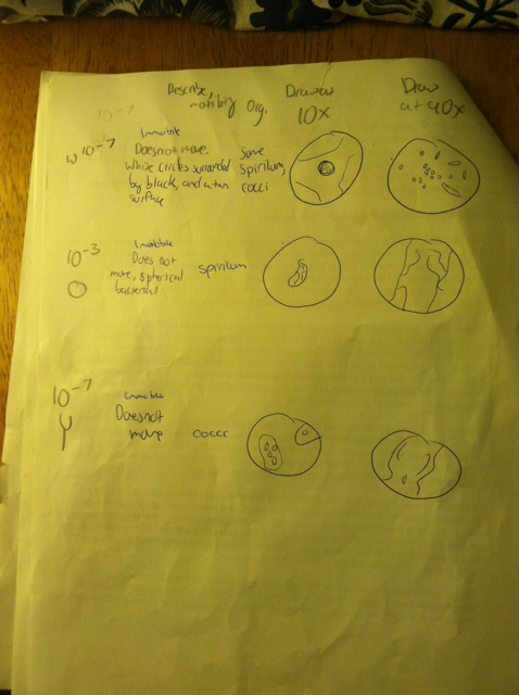
1: Immobile, some spirilum/cocci 2: Immobile, spirilum 3: Immobile, cocci
J.K.
February 9th, 2014
LAB 2: Identifying Algae and Protists
State the question/problem/objective
The point of this lab was to understand how to use a dichotomous key, and to understand the characteristics of algae and protists. I hypothesized that after making observations about the size, shape, movement, and color of single celled organisms the dichotomous key could be used to confirm their identity. In this experiment,the dichotomous key consists of two morphological choices. Starting with the first of two choices, the one that is most accurate for the organism is selected, and then you move to where the choice leads you. Then, there is a second pair of choices. This process repeats until the organism is identified, and diagrams confirm the organism's identity.
1. Wet-mounts from the bottle of 8 protists were made of the Hay-Infusion culture. The gunk from the bottom of the bottle was gathered.
2.The protists were identified using the Dichotomous Key at 4x and 10x. The organisms were also observed with the 40x objective, but they were difficult to see because of the motility.
3.Our Hay-Infusion Cultures were carefully moved to the bench.
4.Observations were made of the Hay-Infusion culture: any changes in smell, organisms, apparent life, etc.
5.Wet-mounts were made from the Hay-Infusion culture, and any protists were identified. 2 Niches were selected and 3 organisms from each niche were identified for a total of 6 organisms. The observed organisms were drawn and recorded in our notebooks.
6.The bacterial prep begun, first by mixing the Hay-Infusion Culture. Serial 100-fold dilutions of the culture were prepared, and inoculated onto agar petri dishes to be observed for next week.
7.Four tubes of 10 mLs sterile broth were obtained and labeled as 2, 4, 6, 8. Four nutrient agar plates and four agar plus tetracycline plates were selected. 8. All the tetracycline plates were labeled with "tet". One plate from each of the two groups were labeled as 10^-3, 10^-5, 10^-7, and 10^-9. Initials were added to identify the group.
9. Then, the Hay Infusion Culture was swirled to mix up all the organisms. 100 mL was taken from the culture and added to 10 mLs for the 10^-2 dilution and swirled. 100 mL of broth from tube 2 was used to inoculate tube 4 and swirled. This process repeated two more times to make the 10^-6 and 10^-8 dilutions.
10.100 microliters was taken from the 10^-2 tube and placed on the nutrient agar plate marked 10^-3 and spread. This process was repeated on the tet plate 10^-3, creating another 10-fold dilution. This procedure repeated with the number 4 tube on the 10^-5 plate, number 6 tube on the 10^-7 plate, and number 8 tube with the 10^-9 plate.
11.The agar plates were incubated at room temperature over the following week.
Shown below
State conclusions and future plans
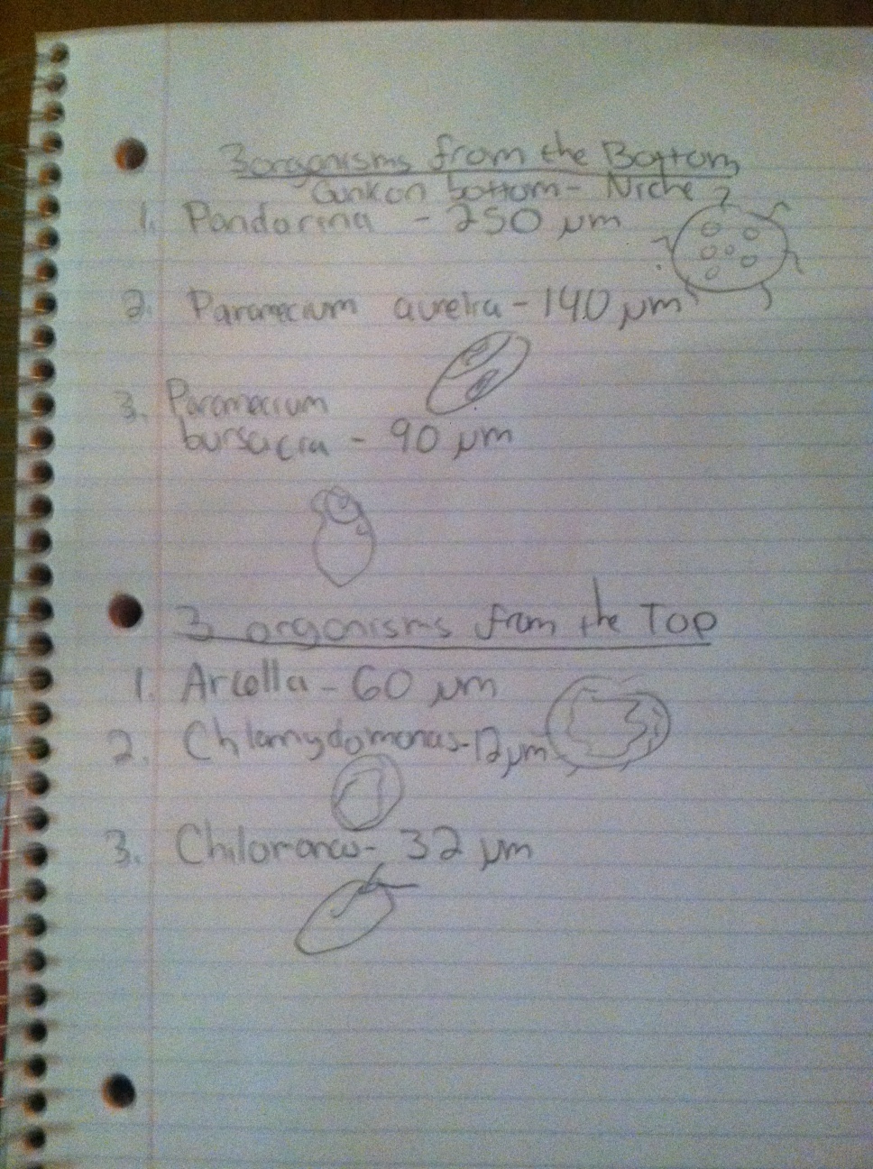
By using the dichotomous key, organisms from the bottom niche and top niche of our culture were properly identified. In the bottom niche, (the gunk on the bottom), pandorina, paramecium aurelia, and paramecium busaria were identified. On the top nice, (top film layer on the water), arcella, chlamydomonas, and chilomonas were identified. By examining the organism’s size, shape, movement, and color, we were able to learn and understand how to use the dichotomous key. Therefore, the objective was explicitly addressed. This also in turn helped us understand and research the characteristics of algae and protists present in the culture. One thing I was surprised about in this lab was that we did not find more photosynthetic organisms on near or on the surface of our culture. Photosynthetic organisms need to live near or on top of water in order for the light to reach them for photosynthesis. There was a clear transformation in our Hay Infusion Culture. The smell of the culture was musky and foul. There was a layer of film that had developed at the top of the jar. There was a thick layer of sludge that was resting at the bottom of the jar, and small spots of green mold.
Organisms near vs away from the plants will different because the species in the area are exposed to two different environments. Species that live far away from the plant may eat the mold that is resting on the bottom of the jar, while species near the plant could feed upon the plant matter.
We got our samples from the top film layer and from the sludge resting at the bottom:
3 Organisms from Bottom Niche- Gunk on Bottom 1. Pandorina- 250 micrometers, green algae, pandorina genus, mobile using flagella. 2. Paramecium Aurelia-140 micrometers, paramecium genus, mobile using cilia. 3. Paramecium Busaria- 90 micrometers, protozoan, paramecium genus, mobile-mobile.
3 Organisms from Top Niche-top film layer of water 1. Arcella- 60 micrometers, Arcella genus, mobile 2. Chlamydomonas- 12 micrometers, green algae, Chlamydomonas genus, mobile with flagella 3. Chilomonas - 32 micrometers, chryptophyte, protozoa, chilomonas genus, mobile with flagella
- these organisms do not photosynthesize
Chosen species: Paramecium Aurelia
A Paramecium is a heterotroph, and it gets its food from nutrients within the hay infusion culture (like algae).Paramecium is a genus of unicellular ciliate protozoa. Paramecium move using their cilia and spiral throughout the water. They use a large amount of energy to propel through water. This paramecium reproduces asexually through a process called binary fission.
If the Hay Infusion Culture was observed for another two months, I would expect to see an increase in the amount of mold/algae, and an increase in the amount of organisms living in it because of an increase in bacteria. The color of the culture would darken as more matter built up, and the smell would become increasingly potent.
There is the selective pressure of the search for food and nutrients for the organisms living in the Hay Infusion culture. They are competing for these resources available, as well as mating with various organisms. For instance, if there is a limited amount of plant matter only the fittest organisms can survive.
J.K
1.31.14
LAB 1
State the question/problem/objective
The objective of this lab was to learn how to properly identify and evaluate the evolutionary specialization of members of the Volvocine Line, while practicing the use of the compound microscope and other techniques such as the addition to protosol. It was also important to recognize and learn how to identify biotic and abiotic characteristics of a niche. I hypothesized that by identifying the diversity among Volvox, Gonium, and Chlamydomonas, Darwin's ideas about natural selection within the field of population genetics would be supported. If the types of green algae exhibit variability, differential capacity for survival and reproduction, and heritable traits, then natural selection is occurring. Variability is demonstrated by aspects such as the number of cell numbers or colony sizes. Then, the reproductive specialization (isogamy vs oogamy) can be evaluated. The heritable traits could be witnessed by varying traits between male and female in the same species. For instance, in Volvox the female gamete is larger than the male. The history of this trend could be researched to test whether or not this trait has successively passed on for females because it leader to higher fitness. This could be logical considering that Volvox are oogamy.
Record the Specific Steps Performed
1. We observed the isogamous, single-celled, motile algae named Chlorophyta. In order to do this, protoslo was added so that the motile creates could be examined more easily. 2.The pyrenoid, stigma, chloropast, and flagella were identified. The flagella was observed by closing the iris diaphragm of the microscope in order to reduce the light. The nucleus was hard to see, because the nucleus is typically hard to see in living cells. 3. In Table 1, the number of cells, colony size, functional specialization of cells, and reproductive specialization was recorded. 4.The living culture and complex species of Gonium was placed onto a slide and observed. 5. Highly complex Volvox was observed. For both Gonium and Volvox, the same characteristics listed for Clamydomonas were then observed and recorded in Table 1. 7. After observing the algae under the microscope, my group was assigned a transect (Transect #5). 8. We proceeded to the transect with zip-lock bags in order to collect samples from our transect, and our notebooks to collect notes.The dimensions of the transect were already defined for us. 9. Once we arrived at our transect, the general characteristics of the transect (location, topograph) and the abiotic and biotic components of the transect were recorded. 10. We collected ground vegetation samples of our transect in zip-lock bags. 11.In order to make a hay infusion culture, 10 grams of the soil and ground sample was placed into a plastic jar containing 500 mLs of deer-park water. 12. 0.1 gm of dried milk was added to the transect, and the entire mixutre was gently mixed for 10 seconds. 13. The top of the jar was removed, and the open jar was placed upon the table in the back of the lab. The jar was labeled so we could identify our Hay Infusion Culture.
Shown below
In Table 1, it can be observed that Chlamydomonas, Gonium, Volvox exhibit diversity in their motility, reproductive qualities, cell and colony sizes. This supports the fact that natural selection is occurring because the species are diverse, they demonstrate differences in their abilities, and they possess different traits that must have passed on due to the fitness values. Therefore, the hypothesis was supported by the data and the objective was explicit. The Chlamydomonas proved to exhibit great diversity, as was discussed in the lab. Our transect possessed both abiotic and biotic components that were identified. In the next lab, we use the Hay Infusion culture to observe protists and to inoculate agar petri plates to study bacteria.
Describe the general characteristics of the transect: location, topography, etc. : The transect was located in a grassy area in the middle of two of three large trees near the Washington Seminary. On either side of the grassy area covered in dirt, leaves, and sticks stood two tall trees. The ground was completely covered in leaves because they had fallen from the large trees. The ground and soil was slightly damp from rainfall. In the middle of these two trees was a picnic table and a small, round tennis ball. We also witnessed a small yellow-crested bird perch upon one of the tall trees.
List the abiotic and biotic components:
Abiotic: Tennis ball, picnic table, soil
Biotic: Trees, leaves, bird
Table 1 Evolutionary Specialization of Members of the Volvocine Line
Characteristic Chlamydomonas Gonium Volvox
Number of cells 2 3 1
Colony size 6 5 1
special function motile still still
reproduction specialization isogamy oogamy oogamy
J.K.


