User:Liam Purdy/Notebook/Biology 210 at AU: Difference between revisions
Liam Purdy (talk | contribs) No edit summary |
Liam Purdy (talk | contribs) No edit summary |
||
| Line 1: | Line 1: | ||
'''February 25, 2015---Present Date''' | '''February 25, 2015---Present Date''' | ||
'''Enter the Zebrafish''' | '''Enter the Zebrafish''' | ||
This lab will be carried out over the next few lab classes. It involves the observations of various effects of chemicals on the growth of Zebrafish. The chemicals used in this group were Fluoride and | This lab will be carried out over the next few lab classes. It involves the observations of various effects of chemicals on the growth of Zebrafish. The chemicals used in this group were Fluoride and Rhodamine. 20 mLs of deer park water were used as a control. The other two dishes contained 20 mL the tested chemical variables. 20 healthy eggs were then put into each of the three petri dishes and allowed to sit for 2 days before being checked on. The fish were fed regularly and dead fish and gunk were consistently removed from the fluid. | ||
-LP | -LP | ||
| Line 12: | Line 12: | ||
Photo 1: | Photo 1: | ||
This photo was taken as I was walking between Bender and Leonard Hall. | This photo was taken as I was walking between Bender and Leonard Hall. It took me 10 minutes, complete silence, and a lot of chasing to get this. | ||
[[Image:IMG 5131 | [[Image:IMG 5131 Cardinal.JPG]] | ||
Based off of the observations of this lab, as well as visual observations from just passing through the transect, a food web is able to be made to show how the organisms rely or feed off one another in order to create a stabilized and functioning ecosystem. | Based off of the observations of this lab, as well as visual observations from just passing through the transect, a food web is able to be made to show how the organisms rely or feed off one another in order to create a stabilized and functioning ecosystem. | ||
Photo 2: | |||
The photo below is a hand drawn food web. This is how I believe the ecosystem functions. | |||
Revision as of 17:40, 4 March 2015
February 25, 2015---Present Date
Enter the Zebrafish
This lab will be carried out over the next few lab classes. It involves the observations of various effects of chemicals on the growth of Zebrafish. The chemicals used in this group were Fluoride and Rhodamine. 20 mLs of deer park water were used as a control. The other two dishes contained 20 mL the tested chemical variables. 20 healthy eggs were then put into each of the three petri dishes and allowed to sit for 2 days before being checked on. The fish were fed regularly and dead fish and gunk were consistently removed from the fluid. -LP
March 4, 2015
Food Web
One great opportunity that was presented through the transect lab over the past few weeks was that it forced attention towards nature and the organisms living within it. After the first interaction with the transect, I found myself connected to it, as though it were my own plot of land that I had to take care of. While walking between Bender Arena and Leonard Hall, I started making sure to change my route so that I would be able to take a look at the condition of my transect, making sure that nothing was torn up or damaged.
The observation labs that were required parts of this lab section also revealed the wide variety or organisms that inhabit the transect. From the larger, more obvious organisms like the birds that are just flying through and the squirrels that inhabit the trees, to the smaller protists and microorganisms that inhabit the fallen leaves of the transect floor, this lab has made it more apparent that there are living creatures everywhere whether visible or not.
Photo 1:
This photo was taken as I was walking between Bender and Leonard Hall. It took me 10 minutes, complete silence, and a lot of chasing to get this.

Based off of the observations of this lab, as well as visual observations from just passing through the transect, a food web is able to be made to show how the organisms rely or feed off one another in order to create a stabilized and functioning ecosystem.
Photo 2: The photo below is a hand drawn food web. This is how I believe the ecosystem functions.
February 19, 2015
Lab 5: Invertebrates
The primary purpose of this laboratory experiment was the become familiar with the different types of invertebrates, as well as to understand their common existence in our transects. This was accomplished by using a Berlese Funnel that would lead to a sample collection of the variety of invertebrates that could be found within our transect. Samples were taken from the top and bottom of the ethanol collection tube, which provided organisms in a variety of sizes.
The setup for the funnel was as follows: First, 25 mL of half ethanol, half water solution was pored into a 50mL conical tube. Next, the collected leaf liter form the transect was placed into a large funnel with a wire screen in the bottom. This screen allowed small organisms to fall out of the funnel, while keeping the plant litter from escaping. The spout of the funnel was then put into the the tube, not touching the solution at the bottom. The two components were then taped together and securely fastened to a stand so that the Berlese Funnel would not tip while being left to sit. A 40 Watt lamp was then placed over the funnel and left on. This would dry out the collection of snow-moistened leaves and would also dry out any tiny invertebrate organisms that were living in the leaf collection. Tin foil was put over the fun land lamp to keep heat concealed.
Photo 1:
The photo below is a hand-drawn rendition of the Berlese Funnel.

The contents of the ethanol were then examined under a dissecting microscope to see what organism samples existed in the litter.
Photo 2:
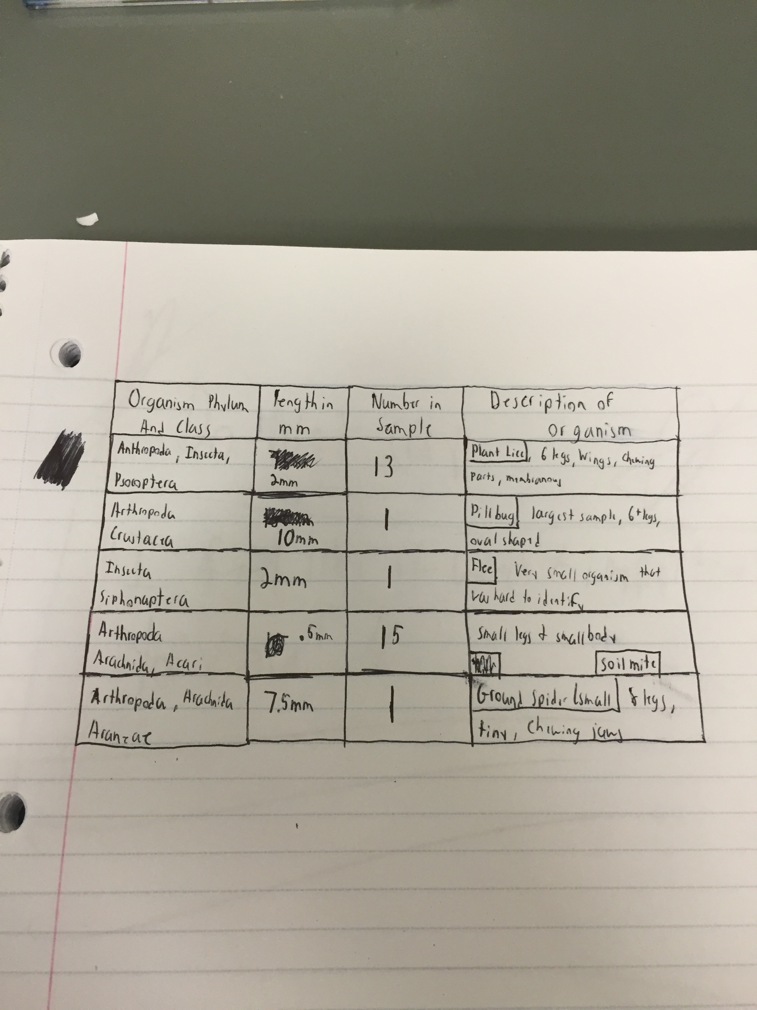
The following photo is of a table from the lab book that recorded the findings of the Berlese Funnel.

Another portion of this lab was the exploration of different types of worms; Acoelomates, Pseudocoelomates, and Coelomates. When I find out how to post a video, I will post a video of the Flatworm Moving. The Flat Worms seemed t slither around their tray in order to move. The Pseudocoelomates moved by wobbling from side to side in a spastic, random motion. Finally, the Annelida earth worms moved like a common worm found in the soil would; scrunching up the mid section in order to propel forward or backwards; this helps the Earth Worm to move through the soil easily. -LP
February 11, 2015
Lab 4: Plantae and Fungi"
The purpose of this lab was to explore the differentiating characteristics between plants and fungi. Another requirement for this lab was to once again get involved with the Transect environment and collect leaf samples for the following week's lab.
The first requirement for this lab was to enter our transect again and collect various leaf samples from the ground and from bushes. The plants form the ground were collected in bulk into a bag, and would be later set up to create a Berlese Funnel. The other leaves would be used to examine the various characteristics and types of leaves in the transect group. The collective details of these plants was summarized in Table 1 of the lab manual.
Photo 1:
This table summarizes the plant collection process and the characteristics of the acquired plants.

Each plant was selected for its uniqueness in comparison to the others. The locations of these plants can be accurately seen by using the hand-drawn map from the first laboratory entrance.
Photo 2:
This plant sample was taken from a tree in the North West portion of the Transect. As you can see, the tree still has numerous green leaves on it, showing that it is able to survive the cold winter months when many trees and other leaf-carrying plants shed their foliage. The leaves grow off the branch in bunches of 5.

Photo 3:
This sample was taken from a low lying, bush-like plant in the southern corner of the transect. These bushes grow all over the floor, covering the whole ground area in a thick, intertwined jungle. The plant is generally on long stalk of leaves on either side.

Photo 4:
This leaf sample was taken from a larger bush that is also located in the Southern corner of the Transect. This medium sized bush still had many of its leaves attached which is why it was selected for examination.

Photo 5:
This sample was taken from a very tall, spiny bush in the South eastern corner of the Transect. Most of the leaves on this one had fallen off. However, it was the fact that this particular bush looked so different from the other samples that it was selected to be observed in this lab.

Photo6:
This final sample was taken from the North East section of the Transect, from a very large bush that still had numerous leaves on it. This bush had lost many of its leaves that were present in the warm weather, but the ones that were retrieved were still green. Each branch, however, only held one or two of these leaves, showing that they were probably about to fall as well.
 -LP
-LP
February 4, 2015
Lab 3: Microbiology
The purpose of the Microbiology lab was the ability examine colonies of bacterium under a microscope, and how to accurately perform tests that allow for identification of the characteristics of a bacteria cell. The test that was to be carried in with this lab was a Gram Stain test, which tests for the existence of peptidoglycan in the bacteria's cel walls; a pink stain is negative and a purple stain was positive. Finally, a PCR for 16S sequencing was also prepared.
The first instructions given during this lab were to retrieve the Hay Culture and to make observations regarding how it has changed since last week. Firstly, the scent of the water, while still terrible, was not as rotten as it had been the week before, although the scent now lingered in the nostrils for a lot longer after smelling. Secondly, there was a drastic decrease in the volume of the water in the ecosystem. What this most likely means is that as more and more bacterial colonies come to exist in the Hay-Infusion, resources will begin to get more and more scarce as competition for survival increases. The water has turned a murkier dark brown and all the leaves have fallen to the bottom of the jar. A final noticeable change in the ecosystem is the presence f a thin, brown and black spotted layer floating on top of and on the walls of the water and jar. This is clearly a new bacterial colony that may require light for nutrients; this would explain why they lie on the top of the water, where light would come to them easiest.
Your notebook is very complete, keep it up! Please try to make your photos smaller, it will be easier for you to use the notebook in the long run. ML
Photo 1:
The following image is a side-view photo of the Transect 3 Hay-Infusion Culture after 2 weeks of sitting.

The next set of observations were based off of the AGAR Nutrient Plates that were prepared last week at the end of the lab period. Each plate contained a different concentration of bacteria, and 4 of the 8 plates were coated in TET, an antibiotic. Bacteria grew on every plate regardless of the presence of the antibiotic. What this indicates is that the bacteria that formed on the TET plate must have had a genetic mutation that granted immunity tot hat particular antibiotic. This resembles a huge issue that is currently plaguing the scientific community today; the release of antibiotics into nature is causing super bacteria that cannot be easily destroyed with conventional medicine. Colonies that grew on the plate ranged in characteristics (other than shape, all were bumped circles). Some colonies were incredibly small while others were very large, some were orange while others were white or a creamy vanilla. One cool growth on the plates was of a white hairy fungus. From my observations, I saw that while there was a significant drop in the number of existent colonies on the plates coated in TET (As shown in picture 3), the plates tended to have larger colonies of bacteria and more fungus growth than than the plates without the TET.
Photo 2:
The image below is of an AGAR plate with bacterial colonies and fungus.

Photo 3:
This is a photo of the 100 Fold Serial Dilution results. This is a chart that records the probable amount of bacterial colonies on the AGAR plate.

The next part of this laboratory experiment was to examine the bacterial colonies under a microscope and to Gram Stain them in order to find the existence of peptidoglycan. In this process, my group chose to examine one colony from four plates, 10^5 TET and no TET, and 10^-3 TET and no TET. Colonies were collected from the plates using a sterile loop and placed onto a wet mount slide. The colony was then looked at through a microscope and different t bacteria and there traits were recorded on a chart (photo 4). Once each plate had an examined colony, the Gram Stain test was prepared. By following the steps that were listed on the paper next to the staining station, we were able to successfully carry out the Gram Stain process. The process of the gram stain included drying out a bacterial colony on a glass slide, then washing over the each slide with a variety of dyes that eventually left the colony with a purple or pink stain, which shows the presence of peptidoglycan. We then examined the slides of bacteria again under the microscope. This time, some of the bacteria had a pink stain, while others had a purple stain. The purple stained bacteria had tested positive for peptidoglycan in its cell walls. The only one that tested positive was the slide from the 10^-3 with TET. This could signify some sort of relationship between high amounts of peptidoglycan and an immunity to certain antibiotics.
Photo 4:
The picture below is a chart that lists the characteristics of the bacterial colonies when examined.

Photo 5:
The picture below a photo of gram negative bacteria through a microscope.

Photo 6:
The photo below is another picture via the microscope of a Gram positive bacteria.

The final part of this lab was to set up a PCR for 16S sequencing. Due to time restraints, my group was only able to make 2 tubes of DNA to be amplified. In the next lab, we will use Agarose gel to find if there is PCR present. If there is, we will be able to purify our DNA for sequencing, which will allow us to identify the bacteria that was present in our ecosystem. The PCR tubes were set up by incubating 100 ul of water and a bacterial colony in 100 C water for 10 minutes. The samples were then centrifuged for 5 minutes while a PCR/ Water tube was prepared. The supernatant was then transferred to the 16S PCR reaction. This was allowed to sit until the next lab. -LP
January 28, 2015
Lab 2: Identifying Algae and Protists
Very complete information so far, great job. Having photos to ID your protists would be helpful. Otherwise, very complete. Keep it up! ML
The main focus of laboratory 2 was to gain the ability to properly identify the differences between protists and algae. In specific, the ability to correctly use a dichotomous key to accurately identify microscopic organisms. Our group was instructed to collect water samples from various areas of our Hay-Infusion ecosystems and analyze the slides under a microscope. We then had to identify the organisms present in each slide using the dichotomous key.
Once our Hay-Infusion was retrieved, the lab book instructed us to analyze how it looked and smelt. The smell was absolutely terrible, resembling only what can be described as an old garbage can left in the sun. The water that was originally clear has turned a disgusting murky green/brown. The soil that we had collected had sunken to the bottom of the mixture and some of the heavier leaves floated in the middle. On top where lighter leaves that had turned a blackish color that showed that they were breeding ground for bacteria. While no visible mold or bacteria were present, on the water, the sides of the jar had gathered a creamy white looking substance that was more than likely mold. Also, the top layer of the jar seemed to have a thick film over it, again probably containing numerous types of bacteria.
Photo 1:
This is a photo of my group's Hay-Infusion after sitting for a week (side view).

Photo 2:
This is a photo of our Hay-Infusion after sitting for a week (top view).

We decided to collect three samples, instead of only two, from the top of the ecosystem, the middle off of a sunken leaf (the leaf had sunken from the top of the Hay-Infusion when the transfer pipette was taking samples from the bottom of the ecosystem. This is why we decided to take a middle sample.), and the bottom from the top of the settled soil. Once each sample was examined, we used the dichotomous key to identify the organisms that we saw. The top sample contained Colpidium (55um), Chlamydoma (9um), and Eudorina (7um). The middle sample contained Colpidium from the top of a sunken leaf. The bottom sample contained Colpidium and Actinospherium (73um). Each of these organisms were motile, which made it slightly difficult to effectively classify them. If our culture was left to grow for 2 more months, and hopefully it will be, I believe that a whole new variety of bacteria would appear, and even more complex ones than the ones that are currently present. I believe that a lot of mold would be present in the jar and in the water, and the water line of the jar would drop drastically. I believe that the leaves would begin to deteriorate from being broken down be the bacteria and the water. Pressure put on the ecosystem would begin to multiply with a drastic increase in the amount of organisms present in the ecosystem. The organisms will have to fight for food and nutrients while also getting the best spots to attract the most light for photosynthesis to occur. The final part of this lab was to create and plate a serial dilution of our Hay Infusion Ecosystem. This was done by first swirling the Hay Infusion, and then removing 100 ul and transferring it to 10 mLs of sterile broth. This created a 10^-2 dilution in this tube. 100 ul of the 10^-2 diluted to a second tube of 10 mL sterile broth; this created a 10^-4 dilution. This process was then repeated two more times for a 10^-6 and 10^-8 dilution. 100 ul of each diluted test tube were then put on two separate agar plates, one with the antibacterial, Tetracycline, and one without it. The diluted mixture was then spread around their respective plates and allowed to grow for the next week.
Photo 3:
This is a hand drawn photo of the serial dilution process:
 -LP
-LP
January 25, 2015 Liam Purdy:
Lab 1: Biological Life at AU
In today's lab, we examined the variations in organisms of the same species that can occur due to evolution. In specific, we examined specimens that were each members of Chlorophyta, a green algae group. The specific line that was examined was the Volvocine Line. Three members of this line were examined in this experiment in order of their evolutionary complexity; Chlamydomonas, Gonium, and Volvox; characteristics of group of organisms were then recorded in the lab book.
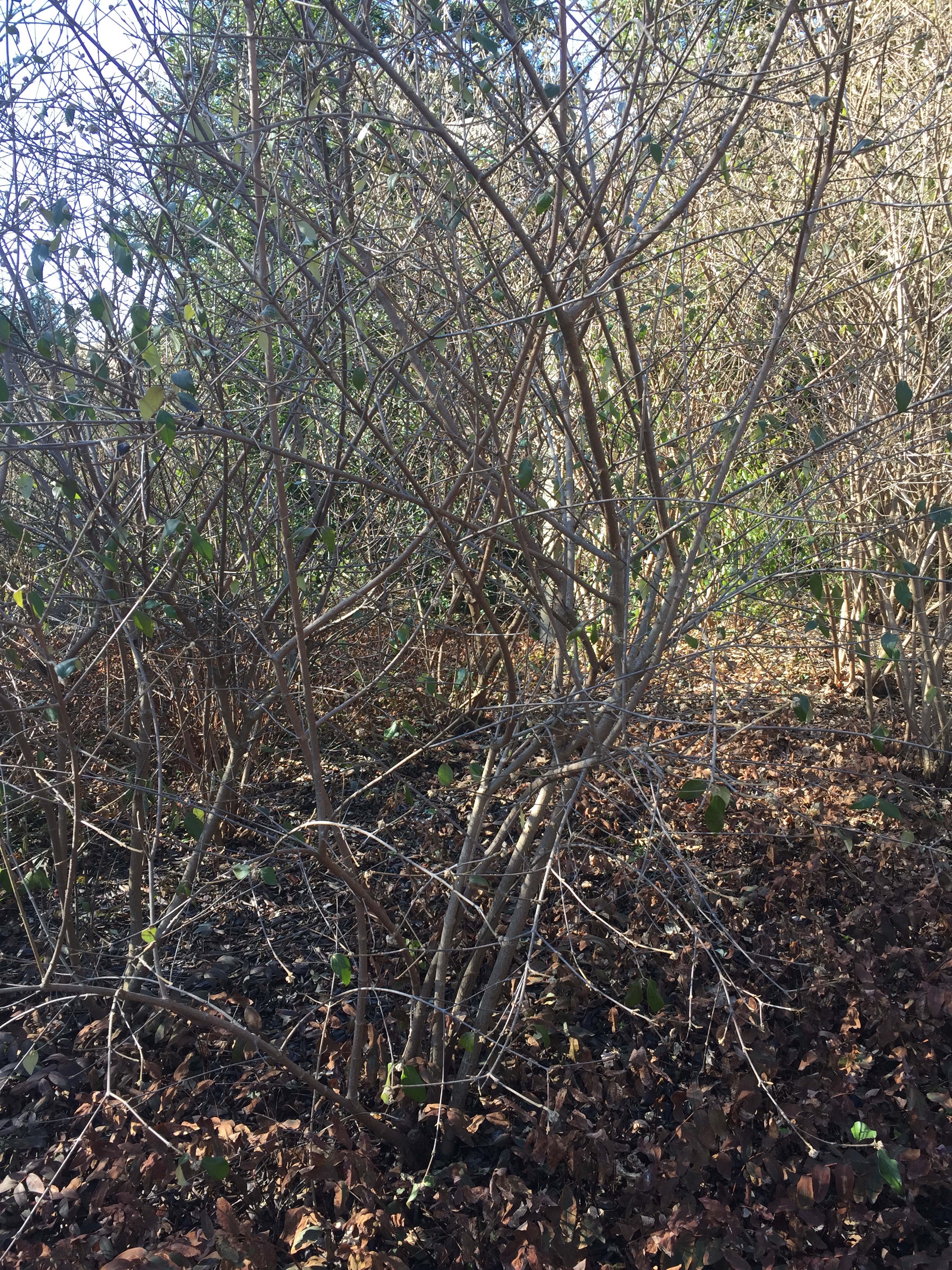
The second portion of this lab was to go outside and examine our group's designated transects. My group received Transect 3, which was located by the bronze AU Eagle statue and referred to as "Tall Bushes". Transect 3 is located right outside of AU's Bender Arena and is the surrounding environment to a small circle with benches for students tot sit and relax in. Because the are has tall foliage, it isn't uncommon for smoking students to enter this secluded area, which in turn means that cigarettes butts are a common cite. The transect is surrounded by concrete sidewalk with more natural features in the center. The middle of this transect is comprised of multiple bushes of various sizes that range from very short, floor hugging bushes to taller thornier bushes. The ground is made of wood chip and dirt, which was relatively frozen due to the recent snow storm.
Photo 1:
This photo is taken from the South corner.

Photo 2:
This photo is taken from the East corner.

Photo 3:
This photo is taken from the North corner

Photo 4:
This photo is taken from the West corner.

Photo 5:
The following image is a hand-drawn arial view of transect 3:

While at our Transect, our group took note of the biotic and abiotic features of the area.
Abiotic Factors: Lampost (2x) Sidewalk/ Concrete Sprinklers (3x) Soil Snow and ice Electrical wires (For the lampposts)
Biotic Factors: Tall and short bushes Wood chips Fallen leaves Low-lying Plants 1 tall tree
In the final part of this lab, each group was instructed to collect various biotic and probiotic specimen from their transects. We were then told to take 10 grams of our collected sample and put it in a jar of 500mL water and .1 g dried milk powder. The mixture was then swirled and let sit or the next week.
Great job compiling all the information and organizing it. Thanks for including the volvocine line information too. Keep up the good work. ML
-LP
January 21, 2015 Liam Purdy. Now I found the way! LP
