User:Lucy Y. Harrelson/Notebook/Biology 210 at AU: Difference between revisions
No edit summary |
No edit summary |
||
| Line 37: | Line 37: | ||
TRICAINE: | TRICAINE: | ||
[[Image:Pic.jpg]] | [[Image:/Users/dejayou1/Desktop/Pic.jpg]] | ||
Revision as of 12:51, 23 March 2014
Instagram: http://instagram.com/bio210farm
Zebra Fish Journal Notes (MARCH): For this zebra fish experiment my lab partner and I were assigned to experiment on the effects of Quantum dots. Quantum dots are nano particles that fluoresce in UV light. the goal was to see how the fish were affected by the nano dots so when later experiments used nano dots, it would be clear what was the new variables affects and what was the nano dots affects. We set up our experiment with three different petri dishes of solutions. Each with 20ml of 0.5 mg/ml of quantum dots, 20ml with 0.05mg/ml quantum dots, and 20ml control with just water. After creating a little habitat to live in, each petri dish of the different solution gets about 20 fish eggs each. We then covered them and let the fish continue to mature for a couple days.
Here are the amount of fish:
Control: 21 fish
0.05: 20 fish
0.5: 20 fish
The first time we checked (about two to three days later) these were the results:
Control: 20 fish
0.05:15 fish
0.5: 17 fish
Then two days later, these were the results:
Control: 16 fish- 2 used for tricaine
0.05: 13 fish-2 used for tricaine
0.5: 15 fish- 2 used for tricane
March 6---> now we use the tricaine fish to do measurements. Tricaine stunts growth so with measurements we are seeing the process of growth. Below is the amount of fish still alive and the charts on measurements of the fish.
Control: all dead fish
0.05: 1 alive fish
0.5: 2 alive fish
TRICAINE:
File:/Users/dejayou1/Desktop/Pic.jpg
Thursday, February 20, 2014: Today we had a condensed laboratory day. We fit in two lab procedures in one class period. The first part was about invertebretes while the other focused on the zebra fish that we will watch grow.
On my instagram there are pictures of the invertebrates that we found in the Berlese funnel. They are also labeled with the type of organism that we identified them as. We found about 5 organisms and viewed them under a dissecting microscope. Using the site below from Hope College, I was able to identify what the different invertebrates in my funnel were.
http://www.hope.edu/academic/biology/leaflitterarthropods/
From our transect, there were many possible animal presences but the organisms that we actually saw were the ones under the microscope. this includes all the pictures of spiders and little bugs that are featured on the instagram. In total we witnessed about 5 different small critters from the Berlese funnel. In the food web made about the transect below, there are the inferred animals included in the web.
Here is a food web based on the organisms observed. They are laid out similar to the Freeman model from page 1150-1151 but instead of lines connecting, if the organism connected to multiple sources of food or eats multiple food supply, the name is listed twice: ARROWS REPRESENT CONSUMPTION
Deer and Rabbit -->Plants
Sparrow (decomposer sometimes) + Robin --> insects and arachnids from transect--> Plants
Sparrow and robin-->plants/seeds
Aracnids--> small insects
Small insects (wasps)-->plants
This was our last look into the transect. There were no more visits post this lab. An overall conclusion from the observations made so far would be that it is not a peak season for our transect's survival. Most of the plants were small growing and little. This could have been because it was still chilly and not an optimal temperature for food garden plants to flourish. The dirt told another story. There were little critters present that had survived the less than optimal temperatures. They help define how our transect operated. The small critters such as wasps showed that there must have been some plant or flower presence that they helped pollinate.
Thursday, February 6: Today we observed plant and fungi given to us. Then we took 5 different plants and fungi from our Farm and tried to classify them. Another note on this lab notebook is that i have made a "Farm" instagram so that i can post picture from there instead of posting them on here. It will be easier to see the compilation of photos from an instagram rather than from a long blog like this. My instagram link is at the top.
The five plants that we found are pictured on my instagram. Here is a table about their info. They are numbered exactly like they are in the picture from instagram. Here is the chart:

Notice that the vascularization varies from plant to plant. Some plants have more vascular qualities to grow taller while others can have structure but are not as sturdy and able to grow upward. Numbers #1, #4, and #5 have broad leaves and are therefore most likely made of dicot seeds. The other two (#2 and #3) may be monocot because of their narrow leaf/pine structure.
Finally, to answer the question about Fungi Sporangia. Fungi Sporangia is the place where spores are formed which helps the fungi reproduce and grow. Since they are asexual, the sporangia is important in their survival.
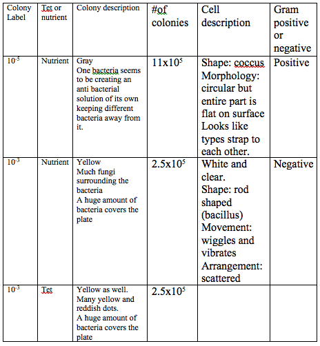
Thursday, January 30, 2014: So after doing this experiment this week We noticed that we had strong colonies in both nutrient and nutrient+tet for 10^-3. So for this weeks labs we did a number of tests on our Farm sample to see what bacteria was living in the area. **Before getting into these test we first re-observed the Hay Infusion Culture from our farm. Nothing really changed. It seemed like there was less water and less of a filament on the water surface.**
-> There seems to be some differences in the colony types between the plates with and without antibiotics. Two of the containers of just nutrients had living bacteria while only 1 of the antibiotic plates had surviving bacteria. The surviving bacteria in each plate looked different. the two 10^-3 plates had surviving yellow bacteria and there was one other nutrient plate (10^-5) that was grey.
-> The effect of tetracycline on the total number of bacteria based on our plates seems to eliminate anything living. The only exception was for 10^-3 with tetracycline which had a good amount of fungi and bacteria. This could be a bacteria that was immune to tetracycline's killing abilities or a possible mix up in plates. Maybe the plate never had tetracycline... that is a possibility and could affect our readings of the results. The only way to be sure would be to redo the plates part of the experiment.
-> The species that are unaffected by tetracycline are staphylococcal (food based infection), streptococcal (strep-throat infection), and pneumococcal (pneumonia/bad cold) infections. They are unaffected because they changed over time to resist the effects of tetracycline.
->Tetracycline works by killing bacteria. It kills a range of bacteria that are detrimental to the respiratory tract. It kills some gram negative and gram positive cells, it kills richettsia and spirochetes bacteria, and can be used on E-coli infections and flu infections.
HERE ARE SOME CHARTS ON DATA FROM BACTERIA (in picture form)-- NOTE Last box is GRAM POSITIVE****
Cites on Tetracycline: http://www.nlm.nih.gov/medlineplus/druginfo/meds/a682098.html http://www.chm.bris.ac.uk/motm/tetracycline/antimicr.htm
Some old pictures related to the Hay Infusion:
Thursday, January 23, 2014: Today we made our hay infusions to see how the bacteria will grow from our sample from the "Farm." The jar with 500 mL of water and the sample of our "Farm" earth.
-- It looks like it has a top layer of fuzz/mini roots growing and hanging down from the top film.
--There is no smell.
--There is dirt at the bottom (light brown).
--There is a middle aqueous layer.
--Has a barren look.
--The soil looks different from when we first put it in. before it was a dark brown and now it looks like an ashy light brown.
-- When looking closely the bottom dirt looked darker than the first layer of dirt exposed to the aqueous layer.
When we looked up our samples top and bottom environments in a microscope this is the data we found:
~Slide for the bottom layer: Observed bacteria were paramecium, 2 gonium, peranema sp that was stuck on debris, vasaria, and diffulgia.
~Slide for top layer: Observed bacteria were gonium, dead paramecium, chlamydomonas, bursaria trancatella, didinium.
Answering Questions: If the hay infusion culture had been observed for another two months, there would be more growth in the jar. There would probably be more time for the bacteria to create more colonies and add to the ecosystem. This means the "Farm" jar would probably look more barren and when taking a sample of it, there would be more bacteria to observe. Some selective pressures that affected the composition were probably the dirt and roots that was in with the bacteria. The big root looks like it contributed to the small root like structures that float on the top and probably affect the flow of oxygen and light. The dirt could have affected the composition since it was so heavy and compacted at the bottom making a place where some underground earth-like bacteria could nuzzle in.
Wednesday, January 22, 2014: I decided to visit the farm. It was FREEZING. I ended up just passing by since it was just too cold and I was too unprepared with my light coat. It is safe to say that snow is covering our plot of land. This means plants are dead/dying, therefore life is probably only visible underneath. I am excited to visit soon after to see if the ice melts or freezes over and to see how that affects the land.
Thursday, January 16, 2014: First Day on Site Today we went to your plot of land that is located in the community garden of AU. There is a birds eye drawing of the area with descriptive labels of all factors we could observe. I plan on posting the picture in the upcoming days. The temperature was chilly. Probably 40 degrees F and was the type of air that is cold to the bone. All plants were either dead or slowly receding as if as if to get away from the chill. No animals in sight. We spotted bunny poop about 15 feet away from our target area which could imply that there are some critters coming in and out of the gated garden. Some Biotic factors: --> wood chips --> plants -->Worms and Critters -->Soil w/ nutrients -->dried roots and lettuce like plants -->Possible bunnies or squirrels in the area as well contributing to the ecosystem Some Abiotic factors: -->Structural wood creating bins for plants -->Rocks -->Sun (a strong presence in the area) -->mesh wire (leftover to keep animals out) -->Plastic plant labels -->paint on the wooden structures to hold the plants in
Basic Info: The Farm is located on AU campus and is better known as the community garden. This notebook is purely for scientific description of the area that I will study over the course of time. My lab partners are Sophie and Amie.
