User:Matthew T. Small/Notebook/Biology 210 at AU: Difference between revisions
No edit summary |
No edit summary |
||
| (20 intermediate revisions by 2 users not shown) | |||
| Line 2: | Line 2: | ||
'''January 26, 2015''' - Lab 1: Biological Life at AU | |||
<br> | |||
<br> <u> Very nice description and aerial view! ML </u> | |||
The transect I received was Transect 5; located on the main campus, right next to the benches and cement patio-like thing in the middle of the quad. Surrounding the patio are bare rose bushes, ferns and a normal bush that was also bare due to the winter. These plants were planted in a planter that went from point D (the northwest corner that was on the edge of the planter) to point B (the southeast corner). Point C was 8.5 steps to the west of point B, so it is in the middle of the planter. Point C is 32 steps from point D; which if you are standing at point C, point D is to your northwest on a diagonal. Point B is 28 steps south of point A, and point A is 24 steps east of point D. The vast majority of the transect was plain grass, with the only non-grass area being the planter. The planter by far had the most components in it. In the planter we found rose bushes, bushes, and ferns (all biotic); we also found soil, snow, rocks, woodchips, cement and leaves (all abiotic). We did find two more biotic features on the grass though; clovers and grass. | The transect I received was Transect 5; located on the main campus, right next to the benches and cement patio-like thing in the middle of the quad. Surrounding the patio are bare rose bushes, ferns and a normal bush that was also bare due to the winter. These plants were planted in a planter that went from point D (the northwest corner that was on the edge of the planter) to point B (the southeast corner). Point C was 8.5 steps to the west of point B, so it is in the middle of the planter. Point C is 32 steps from point D; which if you are standing at point C, point D is to your northwest on a diagonal. Point B is 28 steps south of point A, and point A is 24 steps east of point D. The vast majority of the transect was plain grass, with the only non-grass area being the planter. The planter by far had the most components in it. In the planter we found rose bushes, bushes, and ferns (all biotic); we also found soil, snow, rocks, woodchips, cement and leaves (all abiotic). We did find two more biotic features on the grass though; clovers and grass. | ||
| Line 35: | Line 37: | ||
'''January 27, 2015''' - Lab 2: Identifying Algae and Protists | |||
<br> | |||
<br> <u> Great description of your hay infusion and the organisms. Pictures of the organisms would help me understand what you saw as well as help you remember when you are compiling all this information later. Keep up the good work! ML </u> | |||
The first thing you notice about the culture is the smell; crap, literal crap. It just smells awful. Visually, it looks like everything rose to the top; grass, leaves, etc. There also appears to be a film-like cover formed on the surface of the water. Under this layer of much is yellowish water with strands of grass going through it. Bellow the middle part of the culture, it appears as though all the dirt has fallen to rest at the bottom of the jar. | The first thing you notice about the culture is the smell; crap, literal crap. It just smells awful. Visually, it looks like everything rose to the top; grass, leaves, etc. There also appears to be a film-like cover formed on the surface of the water. Under this layer of much is yellowish water with strands of grass going through it. Bellow the middle part of the culture, it appears as though all the dirt has fallen to rest at the bottom of the jar. | ||
We took two samples from | We took two samples from different niches of our culture; one from the top and one from the bottom of the water. The organisms found on the top of the culture, where plant life was found, would differ with an organism found on the bottom because the organisms on the top will have access to oxygen due to the plants. As a result, the organisms on the bottom would have to find a way to survive without plant life and underwater. | ||
The organisms we found on the top of the culture: | |||
1) This organism was very fast (and therefore motile), colorless, and oval. It moved with the help of cilia all over itself. It was very small, about 37.5μm. We used the dichotomous key to find that it was a Colpidium. It is a protozoa and it does not photosynthesize. | |||
2) This organism was green, flat and was composed of a colony of many cells. We measured it to be 500μm, but that was because there were multiple organisms together. We discovered that this organism is a Gonium. This organism is an algae and also does not photosynthesize. | |||
3) The final organism from the top niche was shaped like a bugle or trumpet and was colorless. It moved in an apparent motion, very fragmented. This was due to it having cilia focused in the back of the organism, so it mainly moved forward or straight. It was measured at about 200μm. We were unable to determine what this organism was. | |||
The organisms we found on the bottom of the culture: | |||
1) This organism was clear and seemed to vibrate. It was covered in cilia and was elongated, measured at 25μm. We determined the organism to be a Spirostomum; a protist who does not photosynthesize | |||
2) This organism had a dark color that wasn't green. It was sperm-like and had a flagella. It was very small, only measuring in at 6-7μm. We were unable to determine this organism as well. | |||
Finally, we were able to see an abundance of Colpidiums and organism 3 from the top niche in the bottom. | |||
In the text, five fundamental characteristics are given as the needs of life. They are; energy, cells, information, replication and evolution. The Spirostomum displays all of these characteristics. | |||
'''Energy'''- Organisms must acquire and use energy to life. Spirostomum uses beating cilia to sweep small organisms into its mouth to eat. | |||
'''Cells'''- Organisms need to be made up of membrane-bound units, or cells. Which a Spirostomum is made up of. | |||
'''Information'''- Organisms need to process information. The nucleus does this for a Spirostomum. | |||
'''Replication'''- Organisms need to replicate themselves. Spirostomums divide in half. | |||
'''Evolution'''- Organisms need to continue to evolve. Recent analyses of ribosomal RNA gene sequences have confirmed that the Spirostomum is a monophyletic group, showing ancestral lineage and evolution. | |||
In two months time, I would not be surprised to see much more life growing in the culture. Some things that would affect the culture could be; leaving the top on the jar (creating an environment with no air), heating or cooling the jar, or destroying part of culture. | |||
Finally, here is a diagram of the serial dilution procedure: | |||
https://richmondschoolbiology.files.wordpress.com/2008/11/serial-dilution.jpg | |||
'''February 2, 2015''' Lab 3: Microbiology and Identifying Bacteria with DNA Sequences | |||
I think that there is no way that Archaea species will grow on agar plates. The environment that we gave the agar plates is not extreme enough for Archaea species to survive. | |||
Our Hay Infusion Culture was basically the same as last week; including the layer of film on the the top of the water. The smell is a lot worse then the previous week though. This may be a result of organisms dying off. The appearance and smell of the culture will change week to week as a result of more organisms being present. | |||
''Table 1: 100-fold Serial Dilution Results'' | |||
'''Dilution. Agar Type. Colonies Counted. Conversion Factor. Colonies/mL''' | |||
10^-3. nutrient. 1,052. x10^3. 1,052,000 | |||
10^-5. nutrient. 2,231. x10^5. 223,100,000 | |||
10^-7. nutrient. 716. x10^7. 7,160,000,000 | |||
10^-9. nutrient. 425. x10^9. 425,000,000, 000 | |||
10^-3. nutrient + tet. 241. x10^3. 241,000 | |||
10^-5. nutrient + tet. 48. x10^5. 48,000,000 | |||
10^-7. nutrient + tet. 12. x10^7. 120,000,000 | |||
10^-9. nutrient + tet. 38. x10^9. 38,000,000,000 | |||
The agar plates with antibiotics present (nutrient + tet) show a drastic decrease in the amount of colonies seen compared to the nutrient agar plate. However, the colonies on the nutrient + tet plates were considerably bigger than the ones on the nutrient agar plate. The impact of tetracycline is quite clear, it nearly decimates the number of colonies in the plate. | |||
Tetracyclines act by binding to the 30S subunit of the ribosome at the A-site. During protein biosynthesis, the new t-RNA with the amino acid attempts to bind to A-site of the ribosome. However, since the A-site is blocked by the tetracycline, the aminoacyl-tRNA cannot bind to it. Thus without the sequential attachment of the tRNA at the A-site, protein biosynthesis cannot occur. By inhibiting protein biosynthesis tetracyclines cause cell death of the bacterial cell (Mehta). The types of bacteria that are sensitive to tetracycline can be seen in this link: http://www.ncbi.nlm.nih.gov/pmc/articles/PMC99026/table/T4/ | |||
''Table 2: Bacteria Characterization'' | |||
'''Colony Label. Plate Type. Colony Description. Cell Description. Gram + or Gram -. Additional Notes''' | |||
Nutrient Yellow. Nutrient. Yellow, Flat, Punctiform. N/A (unable to see). Positive. dilution of 10^-5 | |||
Nutrient White. Nutrient. White, Flat, Punctiform. No movement, black, squiggly edges, Negative. dilution of 10^-5 | |||
large nucleus, spread out. | |||
Tet Yellow. Nutrient+tet. Yellow, Circular, Umbonate. Small, circular, stationary. Pos/Neg. dilution of 10^-7 | |||
. (two types of organisms) | |||
Tet White. Nutrient+tet. White, Circular, Convex. Small, circular, no movement. Post/Neg. dilution of 10^-7 | |||
. (two types of organisms) | |||
''Gram Stain Procedure'' | |||
a. Label slides | |||
b. Heat fix the air dried slide by passing it through a flame three time with the bacterial smear side up | |||
c. Working with a staining tray, cover the smear with crystal violet for 1 minute, then rinse with water | |||
d. Repeat step "c" with Gram's iodine | |||
e. Decolorize by flooding smear with 95% alcohol for 10-20 seconds, rinse | |||
f. Cover the smear with safranin for 20-30 seconds, rinse, and blot excess water | |||
''Procedure IV: Set up PCR for 16S sequencing'' | |||
1. Transfer a single colony of bacteria to 100μL of water in a sterile tube. | |||
2. Incubate at 100°C for 10 minutes in a heat block. Make sure your tubes are floating in water in the heat block. | |||
3. Centrifuge samples for 5 minutes at 13,400 rpm. | |||
4. During the centrifugation, add 20μL of primer/water mixture to a labeled PCR tube. Mix to dissolve the PCR bead | |||
5. Transfer 5μL of supernatant from your centrifuged samples to the 16S PCR reaction. Place the tube in the PCR machine. | |||
Mehta, Akul. "Mechanism of Action of Tetracyclines." PharmaXChangeinfo Mechanism of Action of Tetracyclines Comments. N.p., 27 May 2011. Web. 02 Feb. 2015. | |||
'''February 8, 2015''' - Plants and Fungi | |||
''Transect Sample Plant 1'' - Clover | |||
The clover we found was in the flower bed of our transect; right on the edge of the dirt, where the grass meets the planter. The clover was green with three leaves and a intricate root system that would make it a dicot. In addition, it goes through vascular vascularization. | |||
''Transect Sample Plant 2'' - Rose bush | |||
This plant was a rose bush, with flowers, bulbs and thorns, found in the planter. It had a net-like leaf and is therefore a dicot. With the ability to flower, the rose bush would be classified as an angiosperm. In addition, it goes through vascular vascularization. | |||
''Transect Sample Plant 3'' - Grass | |||
This plant was a grouping of blades of group, connected by dirt and the root system; found on the edge of the planter. Due to their long, narrow leaves, the grass plant would be classified as a monocot. And by reproducing through flowering, is considered an angiosperm. In addition, it goes through vascular vascularization. | |||
''Transect Sample Plant 4'' - Bulb | |||
This bulb was also found on the edge of the planter. It had thick wavy leaves with red stems. The fibrous root system would make it a monocot. In addition, it goes through vascular vascularization. | |||
''Transect Sample Plant 5'' - Fern | |||
The fern leaf was found in the middle of the flower bed, next to a fern bush. It is a single, very long, slightly green leaf. Due to its parallel leaves, it would be considered a monocot. Through the spreading of spores (sexual reproduction), the fern is an angiosperm. In addition, it goes through vascular vascularization. | |||
Fungi sporangia are hydra filaments that grow upward and form black, globelike structures. The sporangia houses the fungi spores, which are the sex cells of the organism. We were instructed to look at the three dissecting microscopes and decide if they were fungi and which of the three groups they belonged too. The first microscope had a slide of a mushroom, a basidiomycota. The second microscope had mold, or ascomycota. Finally, the last microscope had rhizopus, which is apart of the zygomycota group. | |||
<br> | |||
<br><u> Great job, very complete! Having pictures would be really helpful, but your descriptions are very well done. ML </u> | |||
'''February 18, 2015''' Lab 5: Invertebrates | |||
''Procedure I: Acoelomates, Pseudocoelomates, and Coelomates'' | |||
Flatworm - due to the flatworms blobish, flat body structure, the worms movements could be described as floating over the surface. Extremely slow as well. | |||
Nemotoda - the small, snakelike structure of the nemotodas is the reason for the darting movements. The movements can be seen as slithering. | |||
Earthworm - the long, tubular body structure is the prime reason for the earthworm's way of movement. They stretch and constrict. | |||
''Procedure III: Analyzing the Invertebrates Collected with the Berlese Funnel'' | |||
From our Berlese Funnel we found four organisms, two in the top and two in the bottom of the ether: | |||
Bee/Wasp - we found the bee on the the top of the ether. It belongs to the phylum arthropoda and class insecta. It measured at about 2 mm. We only found one bee in our sample. The bee had two pairs of legs on each side, a long antenna and 2 sets of wings. | |||
Earwig - we found the earwig on the the top of the ether. It belongs to the phylum arthropoda and class insecta. It measured at about 40 mm. We only found two earwigs in our sample; one on the top of the sample and one on the bottom. The earwig was pretty big, segmented, and had six legs, no wings, 2 tails and appendages on the abdomen. | |||
Millipede - we found the millipede on the the bottom of the ether. It belongs to the phylum arthropoda and class diplopoda. It measured at about 15 mm. We only found three earwigs in our sample. The earwig had over 8 legs and was extremely long | |||
Cicada hopper - we found the cicada on the the bottom of the ether. It belongs to the phylum arthropoda and class insecta. It measured at about 50 mm. We only found one cicada in our sample. The cicada had four legs and two sets of wings. | |||
The five | |||
Latest revision as of 16:13, 23 February 2015
January 21, 2015 - I made this. MS
January 26, 2015 - Lab 1: Biological Life at AU
Very nice description and aerial view! ML
The transect I received was Transect 5; located on the main campus, right next to the benches and cement patio-like thing in the middle of the quad. Surrounding the patio are bare rose bushes, ferns and a normal bush that was also bare due to the winter. These plants were planted in a planter that went from point D (the northwest corner that was on the edge of the planter) to point B (the southeast corner). Point C was 8.5 steps to the west of point B, so it is in the middle of the planter. Point C is 32 steps from point D; which if you are standing at point C, point D is to your northwest on a diagonal. Point B is 28 steps south of point A, and point A is 24 steps east of point D. The vast majority of the transect was plain grass, with the only non-grass area being the planter. The planter by far had the most components in it. In the planter we found rose bushes, bushes, and ferns (all biotic); we also found soil, snow, rocks, woodchips, cement and leaves (all abiotic). We did find two more biotic features on the grass though; clovers and grass.
http://media-cache-ak0.pinimg.com/736x/0e/e3/e8/0ee3e85c0d64e216c8bc9f8dea337014.jpg Bush Type 1 (rose bush)
http://media-cache-ec0.pinimg.com/736x/99/d9/58/99d958ecd2c5eda6cfd84037d250156f.jpg Bush Type 2
http://media-cache-ec0.pinimg.com/236x/36/bb/3e/36bb3ee84e64602895dd37a6fdee7c49.jpg Soil/Woodchips/Rocks/Snow
http://media-cache-ec0.pinimg.com/236x/06/bc/e8/06bce833869f906555e7bf125f25bdc5.jpg Grass
http://media-cache-ak0.pinimg.com/736x/4b/9b/59/4b9b5979689afe03338016f3beb16fab.jpg Fern
http://media-cache-ak0.pinimg.com/736x/c9/a5/ff/c9a5ff8d6e08c17cedf04d8823a00c28.jpg Corner A looking at Corner B
http://media-cache-ec0.pinimg.com/236x/46/59/b0/4659b0bd56c1f4dc12de24239366dbad.jpg Corner A looking at Corner D
http://media-cache-ak0.pinimg.com/736x/57/97/cb/5797cbc3b03b6cef5c776d72960bfa3b.jpg Corner D looking at Corner A
http://media-cache-ec0.pinimg.com/736x/d9/b9/44/d9b944cd8707d6b696895b3408c41401.jpg Corner C looking at Corner D
January 27, 2015 - Lab 2: Identifying Algae and Protists
Great description of your hay infusion and the organisms. Pictures of the organisms would help me understand what you saw as well as help you remember when you are compiling all this information later. Keep up the good work! ML
The first thing you notice about the culture is the smell; crap, literal crap. It just smells awful. Visually, it looks like everything rose to the top; grass, leaves, etc. There also appears to be a film-like cover formed on the surface of the water. Under this layer of much is yellowish water with strands of grass going through it. Bellow the middle part of the culture, it appears as though all the dirt has fallen to rest at the bottom of the jar.
We took two samples from different niches of our culture; one from the top and one from the bottom of the water. The organisms found on the top of the culture, where plant life was found, would differ with an organism found on the bottom because the organisms on the top will have access to oxygen due to the plants. As a result, the organisms on the bottom would have to find a way to survive without plant life and underwater.
The organisms we found on the top of the culture: 1) This organism was very fast (and therefore motile), colorless, and oval. It moved with the help of cilia all over itself. It was very small, about 37.5μm. We used the dichotomous key to find that it was a Colpidium. It is a protozoa and it does not photosynthesize. 2) This organism was green, flat and was composed of a colony of many cells. We measured it to be 500μm, but that was because there were multiple organisms together. We discovered that this organism is a Gonium. This organism is an algae and also does not photosynthesize. 3) The final organism from the top niche was shaped like a bugle or trumpet and was colorless. It moved in an apparent motion, very fragmented. This was due to it having cilia focused in the back of the organism, so it mainly moved forward or straight. It was measured at about 200μm. We were unable to determine what this organism was.
The organisms we found on the bottom of the culture: 1) This organism was clear and seemed to vibrate. It was covered in cilia and was elongated, measured at 25μm. We determined the organism to be a Spirostomum; a protist who does not photosynthesize 2) This organism had a dark color that wasn't green. It was sperm-like and had a flagella. It was very small, only measuring in at 6-7μm. We were unable to determine this organism as well. Finally, we were able to see an abundance of Colpidiums and organism 3 from the top niche in the bottom.
In the text, five fundamental characteristics are given as the needs of life. They are; energy, cells, information, replication and evolution. The Spirostomum displays all of these characteristics.
Energy- Organisms must acquire and use energy to life. Spirostomum uses beating cilia to sweep small organisms into its mouth to eat.
Cells- Organisms need to be made up of membrane-bound units, or cells. Which a Spirostomum is made up of.
Information- Organisms need to process information. The nucleus does this for a Spirostomum.
Replication- Organisms need to replicate themselves. Spirostomums divide in half.
Evolution- Organisms need to continue to evolve. Recent analyses of ribosomal RNA gene sequences have confirmed that the Spirostomum is a monophyletic group, showing ancestral lineage and evolution.
In two months time, I would not be surprised to see much more life growing in the culture. Some things that would affect the culture could be; leaving the top on the jar (creating an environment with no air), heating or cooling the jar, or destroying part of culture.
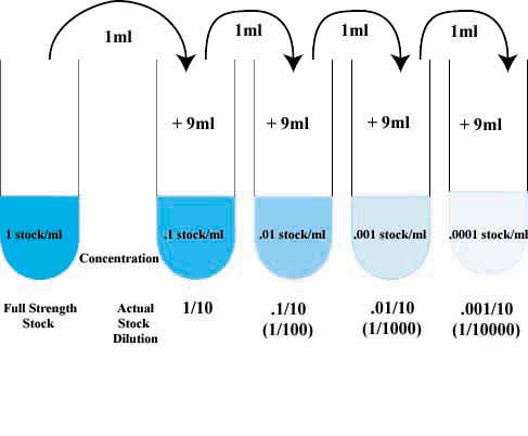
Finally, here is a diagram of the serial dilution procedure:
https://richmondschoolbiology.files.wordpress.com/2008/11/serial-dilution.jpg
February 2, 2015 Lab 3: Microbiology and Identifying Bacteria with DNA Sequences
I think that there is no way that Archaea species will grow on agar plates. The environment that we gave the agar plates is not extreme enough for Archaea species to survive.
Our Hay Infusion Culture was basically the same as last week; including the layer of film on the the top of the water. The smell is a lot worse then the previous week though. This may be a result of organisms dying off. The appearance and smell of the culture will change week to week as a result of more organisms being present.
Table 1: 100-fold Serial Dilution Results
Dilution. Agar Type. Colonies Counted. Conversion Factor. Colonies/mL
10^-3. nutrient. 1,052. x10^3. 1,052,000
10^-5. nutrient. 2,231. x10^5. 223,100,000
10^-7. nutrient. 716. x10^7. 7,160,000,000
10^-9. nutrient. 425. x10^9. 425,000,000, 000
10^-3. nutrient + tet. 241. x10^3. 241,000
10^-5. nutrient + tet. 48. x10^5. 48,000,000
10^-7. nutrient + tet. 12. x10^7. 120,000,000
10^-9. nutrient + tet. 38. x10^9. 38,000,000,000
The agar plates with antibiotics present (nutrient + tet) show a drastic decrease in the amount of colonies seen compared to the nutrient agar plate. However, the colonies on the nutrient + tet plates were considerably bigger than the ones on the nutrient agar plate. The impact of tetracycline is quite clear, it nearly decimates the number of colonies in the plate.
Tetracyclines act by binding to the 30S subunit of the ribosome at the A-site. During protein biosynthesis, the new t-RNA with the amino acid attempts to bind to A-site of the ribosome. However, since the A-site is blocked by the tetracycline, the aminoacyl-tRNA cannot bind to it. Thus without the sequential attachment of the tRNA at the A-site, protein biosynthesis cannot occur. By inhibiting protein biosynthesis tetracyclines cause cell death of the bacterial cell (Mehta). The types of bacteria that are sensitive to tetracycline can be seen in this link: http://www.ncbi.nlm.nih.gov/pmc/articles/PMC99026/table/T4/
Table 2: Bacteria Characterization
Colony Label. Plate Type. Colony Description. Cell Description. Gram + or Gram -. Additional Notes
Nutrient Yellow. Nutrient. Yellow, Flat, Punctiform. N/A (unable to see). Positive. dilution of 10^-5
Nutrient White. Nutrient. White, Flat, Punctiform. No movement, black, squiggly edges, Negative. dilution of 10^-5
large nucleus, spread out.
Tet Yellow. Nutrient+tet. Yellow, Circular, Umbonate. Small, circular, stationary. Pos/Neg. dilution of 10^-7 . (two types of organisms)
Tet White. Nutrient+tet. White, Circular, Convex. Small, circular, no movement. Post/Neg. dilution of 10^-7 . (two types of organisms)
Gram Stain Procedure a. Label slides b. Heat fix the air dried slide by passing it through a flame three time with the bacterial smear side up c. Working with a staining tray, cover the smear with crystal violet for 1 minute, then rinse with water d. Repeat step "c" with Gram's iodine e. Decolorize by flooding smear with 95% alcohol for 10-20 seconds, rinse f. Cover the smear with safranin for 20-30 seconds, rinse, and blot excess water
Procedure IV: Set up PCR for 16S sequencing
1. Transfer a single colony of bacteria to 100μL of water in a sterile tube.
2. Incubate at 100°C for 10 minutes in a heat block. Make sure your tubes are floating in water in the heat block.
3. Centrifuge samples for 5 minutes at 13,400 rpm.
4. During the centrifugation, add 20μL of primer/water mixture to a labeled PCR tube. Mix to dissolve the PCR bead
5. Transfer 5μL of supernatant from your centrifuged samples to the 16S PCR reaction. Place the tube in the PCR machine.
Mehta, Akul. "Mechanism of Action of Tetracyclines." PharmaXChangeinfo Mechanism of Action of Tetracyclines Comments. N.p., 27 May 2011. Web. 02 Feb. 2015.
February 8, 2015 - Plants and Fungi
Transect Sample Plant 1 - Clover The clover we found was in the flower bed of our transect; right on the edge of the dirt, where the grass meets the planter. The clover was green with three leaves and a intricate root system that would make it a dicot. In addition, it goes through vascular vascularization.
Transect Sample Plant 2 - Rose bush
This plant was a rose bush, with flowers, bulbs and thorns, found in the planter. It had a net-like leaf and is therefore a dicot. With the ability to flower, the rose bush would be classified as an angiosperm. In addition, it goes through vascular vascularization.
Transect Sample Plant 3 - Grass
This plant was a grouping of blades of group, connected by dirt and the root system; found on the edge of the planter. Due to their long, narrow leaves, the grass plant would be classified as a monocot. And by reproducing through flowering, is considered an angiosperm. In addition, it goes through vascular vascularization.
Transect Sample Plant 4 - Bulb This bulb was also found on the edge of the planter. It had thick wavy leaves with red stems. The fibrous root system would make it a monocot. In addition, it goes through vascular vascularization.
Transect Sample Plant 5 - Fern The fern leaf was found in the middle of the flower bed, next to a fern bush. It is a single, very long, slightly green leaf. Due to its parallel leaves, it would be considered a monocot. Through the spreading of spores (sexual reproduction), the fern is an angiosperm. In addition, it goes through vascular vascularization.
Fungi sporangia are hydra filaments that grow upward and form black, globelike structures. The sporangia houses the fungi spores, which are the sex cells of the organism. We were instructed to look at the three dissecting microscopes and decide if they were fungi and which of the three groups they belonged too. The first microscope had a slide of a mushroom, a basidiomycota. The second microscope had mold, or ascomycota. Finally, the last microscope had rhizopus, which is apart of the zygomycota group.
Great job, very complete! Having pictures would be really helpful, but your descriptions are very well done. ML
February 18, 2015 Lab 5: Invertebrates
Procedure I: Acoelomates, Pseudocoelomates, and Coelomates
Flatworm - due to the flatworms blobish, flat body structure, the worms movements could be described as floating over the surface. Extremely slow as well.
Nemotoda - the small, snakelike structure of the nemotodas is the reason for the darting movements. The movements can be seen as slithering.
Earthworm - the long, tubular body structure is the prime reason for the earthworm's way of movement. They stretch and constrict.
Procedure III: Analyzing the Invertebrates Collected with the Berlese Funnel
From our Berlese Funnel we found four organisms, two in the top and two in the bottom of the ether:
Bee/Wasp - we found the bee on the the top of the ether. It belongs to the phylum arthropoda and class insecta. It measured at about 2 mm. We only found one bee in our sample. The bee had two pairs of legs on each side, a long antenna and 2 sets of wings.
Earwig - we found the earwig on the the top of the ether. It belongs to the phylum arthropoda and class insecta. It measured at about 40 mm. We only found two earwigs in our sample; one on the top of the sample and one on the bottom. The earwig was pretty big, segmented, and had six legs, no wings, 2 tails and appendages on the abdomen.
Millipede - we found the millipede on the the bottom of the ether. It belongs to the phylum arthropoda and class diplopoda. It measured at about 15 mm. We only found three earwigs in our sample. The earwig had over 8 legs and was extremely long
Cicada hopper - we found the cicada on the the bottom of the ether. It belongs to the phylum arthropoda and class insecta. It measured at about 50 mm. We only found one cicada in our sample. The cicada had four legs and two sets of wings.
The five