User:Rachel Scalzo/Notebook/Biology 210 at AU: Difference between revisions
No edit summary |
No edit summary |
||
| Line 1: | Line 1: | ||
4/23/14 | |||
Lab 6- Effect of alcohol on zebrafish development | |||
'''Objectives & Question''' | |||
The purpose of this experiment was to see how various aspects impacted development in organisms. For this specific experiment, we used zebra fish embryos due to the fact that have similar embryonic development to humans, as well as the fact that their shells are transparent. In this experiment, twenty zebra fish developed in a 1.5% of ethanol. | |||
The hypothesis of this experiment is that zebra fish are the ideal organism for studying fetal alcohol syndrome (FAS). The prediction was that the zebra fish that were exposed to ethanol would exhibit similar symptoms to human fetuses exposed to ethanol. | |||
'''Procedure Performed''' | |||
1. Forty zebra fish embryos were gathered and split into half. Twenty would develop in Deer Park Spring water, whereas the other twenty would develop in a 1.5% solution of ethanol. | |||
2. These embryos were observed four times over the course of two weeks, and notes were taken of how many were still alive, what specific features had developed, and any abnormalities were observed. | |||
3. On day 7, three embryos were to be fixated in their current state so that they could be observed the following week. | |||
4. On day 14, final data was collected on how many organisms were still viable, and were compared to the zebra fish that were fixated the previous week. | |||
*Note, step 3 was not performed, so there was no fixated zebra fish observed, or compared to the zebra fish on day 14. | |||
'''Data''' | |||
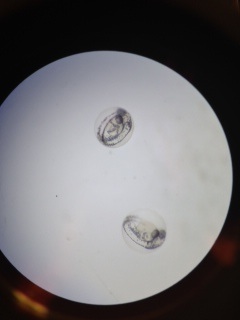
[[Image:Fish, treated, day 1.jpg]] | |||
The picture above shows zebra fish embryos that were treated with ethanol on day 1 of observations. | |||
Observations of untreated, day 1: | |||
-Eyes have developed | |||
-Visible traces of vertebrate column development | |||
-Eggs have black flecks on outer shell | |||
[[Image:Fish, untreated day 1.jpg]] | |||
The picture above shows zebra fish embryos that were not treated and developing in Deer Park Spring water. | |||
Observations of treated, day 1: | |||
-Most have eyes developed, however all eggs have black eye sockets where the eyes will develop | |||
-Eyes not as dark or developed as untreated embryos | |||
-Less black flecks observed | |||
-Spine/vertebrate column shows sign of development | |||
[[Image:Results from first week.jpeg]] | |||
This table represents the observations between the two groups, and comparing them against each other. These observations were taken during the first week of the experiment. | |||
Observations of untreated, day 7: | |||
-Embryos seem to be at pec. fin developmental phase | |||
-No deaths | |||
-Eyes have yellow hue | |||
-Relatively normal in size | |||
Observations of treated, day 7: | |||
-Around long pec. fin phase | |||
-ONe death at larvae stage | |||
-Eyes seem larger in size than untreated embryos | |||
-Have rounder body than average embryo | |||
-Have green/yellow hue to the body | |||
Observations of both groups, day 14: | |||
-All zebra fish in the experiment died | |||
'''Conclusion & Future Plans''' | |||
Based on the data gathered from this experiment, it can be concluded that zebra fish are model organisms to study the effects of FAS, and how those symptoms show on human fetuses. | |||
One suggestion for if this experiment was repeated would be to increase the size of the groups of zebra fish because the larger the sample pool, the more accurate the results would be. Also to feed them around day 8 because then they will not die of starvation. | |||
'''2/24/14''' | '''2/24/14''' | ||
Lab 5- Invertebrates | Lab 5- Invertebrates | ||
Revision as of 15:32, 23 April 2014
4/23/14 Lab 6- Effect of alcohol on zebrafish development
Objectives & Question The purpose of this experiment was to see how various aspects impacted development in organisms. For this specific experiment, we used zebra fish embryos due to the fact that have similar embryonic development to humans, as well as the fact that their shells are transparent. In this experiment, twenty zebra fish developed in a 1.5% of ethanol. The hypothesis of this experiment is that zebra fish are the ideal organism for studying fetal alcohol syndrome (FAS). The prediction was that the zebra fish that were exposed to ethanol would exhibit similar symptoms to human fetuses exposed to ethanol.
Procedure Performed 1. Forty zebra fish embryos were gathered and split into half. Twenty would develop in Deer Park Spring water, whereas the other twenty would develop in a 1.5% solution of ethanol. 2. These embryos were observed four times over the course of two weeks, and notes were taken of how many were still alive, what specific features had developed, and any abnormalities were observed. 3. On day 7, three embryos were to be fixated in their current state so that they could be observed the following week. 4. On day 14, final data was collected on how many organisms were still viable, and were compared to the zebra fish that were fixated the previous week.
- Note, step 3 was not performed, so there was no fixated zebra fish observed, or compared to the zebra fish on day 14.
Data
 The picture above shows zebra fish embryos that were treated with ethanol on day 1 of observations.
Observations of untreated, day 1: -Eyes have developed -Visible traces of vertebrate column development -Eggs have black flecks on outer shell
 The picture above shows zebra fish embryos that were not treated and developing in Deer Park Spring water.
Observations of treated, day 1: -Most have eyes developed, however all eggs have black eye sockets where the eyes will develop -Eyes not as dark or developed as untreated embryos -Less black flecks observed -Spine/vertebrate column shows sign of development
This table represents the observations between the two groups, and comparing them against each other. These observations were taken during the first week of the experiment.
Observations of untreated, day 7: -Embryos seem to be at pec. fin developmental phase -No deaths -Eyes have yellow hue -Relatively normal in size
Observations of treated, day 7: -Around long pec. fin phase -ONe death at larvae stage -Eyes seem larger in size than untreated embryos -Have rounder body than average embryo -Have green/yellow hue to the body
Observations of both groups, day 14: -All zebra fish in the experiment died
Conclusion & Future Plans Based on the data gathered from this experiment, it can be concluded that zebra fish are model organisms to study the effects of FAS, and how those symptoms show on human fetuses. One suggestion for if this experiment was repeated would be to increase the size of the groups of zebra fish because the larger the sample pool, the more accurate the results would be. Also to feed them around day 8 because then they will not die of starvation.
2/24/14
Lab 5- Invertebrates
Movement of Worms 1. Coelomates- they move using muscle contractions throughout their entire body. This movement appears to be complicated since the earthworm (coelomate under observation) only has a couple of muscles in total. Based on their movement, it appears as though there are a couple of muscles for elongation- to stretch from one location to another- and a few for muscle contraction- to return the earthworm to its original length.
2. Acoelomates (platyhelmeathes)- these organisms glide across the petri dish by moving their bodies in a side-to-side motion. Since the structure of the acoelomates seems less complex than the coelomates, it also appears as though they have less muscle devoted to movement.
3. Pseudocoelomates (nematoda)- these organisms shimmy/wiggle in order to get from one location to another. This can be seen by comparing the movements of the other two groups, and how they move.The pseudocoelomates have more complex movement than the acoelomates, but they have less muscles than the coelomates.
- Note: we were unable to find 10 different organisms from the berlese funnel. This may be impacted due to the time of collection, which was when many organisms were not active, or away from the transect due to weather.
Based on the measurements gathered, the largest organism that was found was the arachnid (which was 5 mm long). This is a big contrast to the smallest organism found, which was the fly at 0.5 mm. Based on the other organisms observed, it seems that the most common type found in this transect, during the winter, would be small insects that hide among the leaves and underneath the dirt.
A few types of vertebrates that may live in the transect are: -Sparrows (chordata, aves, passeriformes, passeridae, passer domesticus) : sparrows are constantly looking for material to build their nests in the spring. So it would not be uncommon for a sparrow to come to the transect to grab a twig or small branch for building. They also eat invertebrates such as worms and insects that live in the transect. -Squirrels (Chordata, Mammalia, Rodentia, Sciuridae, Sciurus, Sciurus carolinensis) : squirrels are often scavenging for food to eat, so it would not be uncommon to see a squirrel scurrying through the transect in search of some nuts or other food to eat. -Mice (Chordata, Mammalia, Rodentia, Cricetidae, Peromyscus, Peromyscus boylii): mice are always on the move and could live in the transect underneath the plants and dirt. They could also eat the plants that grow during the warmer seasons and hide from predators. -Robins (Chordata, Aves, Passeriformes, Turdidae, Turdus, T. migratorius): like sparrows, robins can also build their nests from materials, like twigs or small branches, found in the transect. They also eat worms and other invertebrates that live in the transect. -Chipmunks (Chordata, Mammalia, Rodentia, Sciuridae, Tamias, Tamias striatus): chipmunks are very similar to squirrels in that they are able to eat some of the plant life that grows in the transect. The foliege also provides shelter and a hiding place from predators.
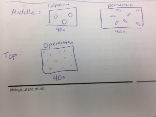
- Sorry, I'm not the best artist.
RS
2/21/14
Lab 4- Plantae and Fungi
Transect 4 Plants
Location: plant #1 was found in a planting box was an angiosperm plant. Description: plant #1 has long leaves that are slightly droopy. The leaves had little buds on the surface that felt soft to the touch. The stem of the plant was very hard/strong wood. Vascularization: Dicot. Leaves and special characteristics: stomata, mesophyll, guard cells, and parenchyma. Seeds, evidence of flowers or other reproductive parts: no evidence was seen or observed.
Location: Plant #2 was found in a planting box and is an angiosperm plant. Description: plant #2 has big, green leaves in groups of three. Is most likely came from a small bush. The edges of the leaves are slightly spiky and are brown compared to the rest of the leaf. Vascularization: dicot. Leaves and special characteristics: the leaves have a waxy type surface. They grow in threes and are big in size. Seeds, evidence of flowers or other reproductive parts: there were no seeds or flowers observed.
Location: Plant #3 was found in the gardening box and was a pteridophyta. Description: plant #3 has long, skinny roots, and fern-like green leaves that are turning brown at the tips. Vascularization: dicot. Leaves and special characteristics: cuticles and stomata. Seeds, evidence of flowers or other reproductive parts: no seeds or flowers were observed.
Location: plant #4 was found in the gardening box and was an angiosperm. Description: plant #4 was short, but was spread in a wide surface area on the ground. It has small, round leaves. Vascularization: dicot. Leaves and special characteristics: leaves are small and round. The stem is weak but has multiple leaves sprouting from it. The majority of the leaves were green, however, some of them were turning a light green/yellow hue. Seeds, evidence of flowers or other reproductive parts: no seeds or flowers were observed.
Location: plant #5 was found in the gardening box and was an angiosperm. Description: It has a short stem, and dark green, oval leaves growing from it. It looks like a weed. Vascularization: Dicot. Leaves and special characteristics: stomata, guard cells and cuticles. The leaves are an oval shape and are dark green color. Seeds, evidence of flowers or other reproductive parts: no seeds or flowers were observed.
- Note: The time of year the plants were all collected was very cold, so none of the plants were displaying any signs of flowering or seeds. The assumption was made that had the time of collection been during a warmer part of the year, then there would have been some indications of flowering or seeds.
-Sporangia grow on the hyphae and are black, spheres that have spores inside of them. The spores are used for when the fungi want to reproduce (which they do asexually). -We were not able to observe any fungi on the agar plates.
RS
2/20/14 Lab 3- Microbiology and Identifying Bacteria with DNA
-I believe that it is possible for archaea to grow on the agar plates, however, since the majority of species of archaea live in extreme environments, the likelihood of archaea growing on the agar plates are slim. Since archaea are able to survive in extreme environments, living on a controlled environment (like the agar plates) should not be problematic for archaea.
Update on Hay Infusion Culture The appearance of the hay infusion would change on a consistent basis due to the fact while new growths and organisms are emerging in the infusion, the debris (such as leaves) are decomposing. This ultimately makes the water turn a murky brown color, and an unpleasant odor to come from the infusion.
Table 1: 100-fold Serial Dilution Results Dilution (Plate Label): 10^-3, nutrient agar, lawn, x 10^3 conversion factor, to many to count ( x>1000) Dilution (Plate label): 10^-5, nutrient agar, 180 colonies, x 10^5 conversion factor, 1.80x10^-5 Dilution (Plate Label): 10^-7, nutrient agar, 69 colonies, x10^7 conversion factor, 69x10^-5 Dilution (Plate label): 10^-9, nutrient agar, 19 colonies, x10^9 conversion factor, 19x10^-9 Dilution (Plate Label): 10^-3, nutrient + tet agar, 53 colonies, x10^3 conversion factor, 52x10^-3 Dilution (Plate Label): 10^-5, nutrient + tet agar, 4 colonies, x10^5 conversion factor, 4x10^-5 Dilution (Plate label): 10^-7, nutrient + tet agar, 0 colonies, x10^7 conversion factor, 0 Dilution (Plate Label): 10^-9, nutrient + tet agar, N/A, x10^9 conversion factor, N/A
-There were a few differences between the colonies that grew on the plates with the tet, and the ones that did not. One difference was that the size of the colonies were smaller on the agar plate with tet than on the plates without. Another aspect that varied between the two different types of plates was the color of plates. The colonies that grew on the agar plates grew in a variety of colors (such as orange, yellow, and pink) whereas the colonies that grew on the tet. plate were mainly orange colonies.
-The different sizes of the colonies show that the presence of tet. on the agar plate impact the size of the colony growth. The overall size of the colony was bigger on the regular tet plate than compared to the tet plate. This shows that the bacteria were able to survive and flourish on the plate without the tet than the plate with the tet. The differing sizes also show that only certain types of bacteria are able to grow on an agar plate with tet on it. For example, the bacteria that form an orange colony are better suited to thrive on the tet plate, than the bacteria that form the red or yellow colonies.
-The presence of tetracycline decreases the overall number/colonies of bacteria that are able to form on the agar plate. The highest number of colonies found on a tet. plate was 53. Compared to the highest number of colonies found on a regular agar plate (which was a full lawn), it is clear to see that the presence of tetracycline decreases the amount of colonies that are able to form. There was no fungi found on any of the agar plates.
-From the observations, it is safe to say that two species of bacteria are unaffected by the tetracycline.
-Tetracycline is an antibiotic used to treat diseases such as pneumonia, acne, and other bacterial infections. Tetracycline stops the growth and spread of bacteria by inhibiting the tRNA from binding to their proteins, ultimately stopping translation from occurring within the bacteria. It is effective on a wide range of bacteria, including many gram positive and gram negative bacteria, Helicobacter pylori (which causes stomach ulcers), and diseases caused by Escherichia coli and Haemophilus influenzae. (MedlinePlus, AHFS, 2014), (Klajn, Chemistry and chemical biology of tetracyclines, 2013).
Table 2 Gram Stain (+), Colony Label #1 (Pink Circle), Tet (-), Pink Circle, 2 colonies of this type, 2x10^-9 bacteria/mL, small pill shaped, often in doubles. Gram Stain (+), Colony Label #2 (Yellow Circle), Tet (-), Yellow Circle, 6 colonies of this type, 6x10^3 bacteria/mL, small, irregular, roundish shape. Gram Stain (-), Colony Label #3 (Orange Circle), Tet (+), Orange Circle, 8 colonies of this type, 8x10^3 bacteria/mL, pill shaped.
References: Klajn, Rafal, (2013). Antimicrobial properties, Chemistry and chemical biology of tetracyclines. Retrieved from http://www.chm.bris.ac.uk/motm/tetracycline/antimicr.htm The American Society of Health-System Pharmacists, Inc., (2014). Tetracycline. Retrieved from http://www.nlm.nih.gov/medlineplus/druginfo/meds/a682098.html
RS
2/9/14
Lab 2- Identifying Algae and Protists
Description of Hay Infusion Culture' The culture smells unpleasant, with a slight odor similar to mold. There are several dark solids at the bottom of the culture, and on the top there are little areas of black mold.
The organisms living in the culture might differ in location (near or away from plants) due to adaptations that they might have developed over the generations. Some organisms have traits better suited to live away from vegetation, for example the food they eat gather on the surface of the water,. Other organisms who live closer to vegetation might need the plants for food, or thrive on less sunlight in the deeper waters.
Organisms observed from the Culture Area 1- The Surface of the Culture 1. Peranema, mobile, protist, not photosynthesizing, 50 um 2. Euglena, mobile, protist, can synthesize, 40 um 3. Actinoshpaerium, mobile, protists, not photosynthesizing, 75 um
Area 2- The Middle of the Culture 1. Peranema, mobile, protist, not photosynthesizing, 50 um 2. Colpidium, mobile, protist, not photosynthesizing, 60 um.
Area 3- Actinoshpaerium, mobile, protist, not photosynthesizing, 75 um.
- Note: we were not able to find 6 different organisms in our culture sample. Several organisms that were found living at the surface of the culture, were also able to survive at the bottom as well.
How Euglena meets the characteristics of life
-Energy: Euglena can get their energy via photosynthesis, or from feeding on food particles, like a heterotroph. Several species of Euglena have chloroplasts so they can take in energy from the sun's rays, however, when there is not an adequate amount of sunlight they can take in food as other heterotrophs do.
- Cells: Euglena are generally unicellular.
- Information: Euglena are able to take in information from their surroundings and adapt to the changes that the environment constantly goes through. For example, Euglena have an organelle called a red eyespot. This organelle is able to filter out certain wavelengths, and in turn, the Euglena is able to turn towards a light source and move to it.
-Replication: Euglena (like most unicellular organisms), reproduce via binary fission.
- Evolution: Euglena is subject to natural selection and evolution due to its' ability to have mutations in its' DNA, and its' ability to adapt to the changes in the environment.
If the Hay Infusion was Observed for Another two Months If they hay infussion was observed for another two months, I predict that there would be several changes, including more mold spores on the surface of the water, denser solids at the bottom of the jar as the mass settles at the bottom, less leaves/debris due to decomposition, and less organisms in the jar. The last aspect would probably be observed due to the decreasing amount of nutrients in the jar over time.
Selective Pressures There were several aspects that affected our sample of the organisms. These included, lack of living space, limited food sources (ultimately leading to predation), and different temperatures from the top and bottom of the hay infusion. The few organisms that were able to survive these pressures had the best ability to adapt to the environment, and survive.
Good, but don't forget the last item in red about the serial dilution. 2/18/14 GHH
1/31/14
Lab 1- Biological Life at AU
Characteristics of transect: Transect #4 is located by the tennis courts at AU. It is contained in a fenced off area, and represents the “farmland”. It is a 20 x 20 ft square that has six wooden boxes for growing plants, which are built on flat ground. The ground is mostly composed of soil and other vegetation.
Biotic Features: Insects, Weeds, Green-Leafy plants, vines, leaves Abiotic Features: Chicken wire, wooden boxes, rocks, dirt, and plastic.
RS


