User:Sithara Thalluri/Notebook/Biology 210 at AU
PCR DATA
>MB47-For_16S_G06.ab1 NNNNNNNNNNNNNNNNNCTNNNCNTGCAGCCGAGCGGTAGAGATTCTTCGGAATCTTGAGAGCGGCGCACGGGTGCGGAA CACGTGTGCAACCTGCCTTTATCAGGGGAATAGCCTTTCGAAAGGAAGATTAATGCCCCATAATATATCATATGGCATCA TTTGATATTGAAAACTCCGGTGGATAAAGATGGGCACGCGCAGGATTAGATAGTTGGTAGGGTAACGGCCTACCAAGTCA GCGATCCTTAGGGGGCCTGAGAGGGTGATCCCCCACACTGGTACTGAGACACGGACCAGACTCCTACGGGAGGCAGCAGT GAGGAATATTGGACAATGGGTGAGAGCCTGATCCAGCCATCCCGCGTGAAGGACGACGGCCCTATGGGTTGTAAACTTCT TTTGTATAGGGATAAACCTACCCTCGTGAGGGTAGCTGAAGGTACTATACGAATAAGCACCGGCTAACTCCGTGCCAGCA GCCGCGGTAATACGGAGGGTGCAAGCGTTATCCGGATTTATTGGGTTTAAAGGGTCCGTANGCTGATGTGTAANTCANTG GTGAAATCTCACANCTTANCTGTGAAACTGCCNTTGATACTGCATGTCTTGAGTGTTGTTGAANTANCTGGAATAANTNN GTANCAGTGAAATGCCTANATATTACTTNNANCACNANGTGCTAANGCANGTTGGTANNCCNCNACTGACNCTGATNGAG NAAANCNTGGGNNAGCGAACANAANTNNATACCCTGGGGNNGTNNNCNNNAANNAANCTNANTCNNTTTTTNTCTTTCTC TTNCNNATACANNNNNANCCGANAAGNTNGCCNNCTNCCGGGTGGTGTTCTCCNTNNTNNNGATGNNNTCNNCTNNNNNN NNNNNCNGCCCCCCCNCAANNATTTNTANANNNNTATANNNTNNNANANCNNGCGGCCCCCTNTNTAANNGGNNNNGGGG GAGNNNNNNGNNNNNGTTTTCTATTATATNTNNNNCTNTNNNCCNCNNGNNCNGGGGGGGTTGTNTCTCCCNNCCAGAAC NNAANGANANTNTNCNNCANCAGCCNNNN
>MB48-For_16S_H06.ab1 NNNNNNNNNNNNNNGCNNANNNTGNNANNNNNGCGGTANGANGGGANGCTTGCTCTNNGATTCAGCGGCGGACGGGTGAG TAATGCCTAGGAATCTGCCTGGTAGTGGGGGACAACGTTTCGAAAGGAACGCTAATACCGCATACGTCCTACGGGAGAAA GCAGGGGACCTTCGGGCCTTGCGCTATCAGATGAGCCTAGGTCGGATTAGCTAGTTGGTGAGGTAATGGCTCACCAAGGC GACGATCCGTAACTGGTCTGAGAGGATGATCAGTCACACTGGAACTGAGACACGGTCCAGACTCCTACGGGAGGCAGCAG TGGGGAATATTGGACAATGGGCGAAAGCCTGATCCAGCCATGCCGCGTGTGTGAAGAAGGTCTTCGGATTGTAAAGCACT TTAAGTTGGGAGGAAGGGCATTAACCTAATACCTTGGTGTTTTGACGTTACCGACAGAATAAGCACCGGCTAACTCTGTG CCAGCAGCCGCGGTAATACAGAGGGTGCAAGCGTTAATCGGAATTACTGGGCGTAAAGCGCGCGTANGTGGTTTGTTAAG TTGGATGTGAAAGCCCCGGGCTCAACCTGGGAACTGCNTTCNAAACTGNCNAGCTAGAGTATGGTANAGGGTGGTGGAAT TTCCTGTGTAGCNNTGAAATGCGTAGATATANGAANGAACACNNNGTGGCNAAGCGGACCACCTGNACTGATACTNACAC TGANNTGCGAAANNNTGTGGANCAAACANNATTANGATNNCCTNNAGTCCACNGCCNGTANACNNNNTCAACTANCNNNN NNAGCNCTTNANNTGTTANTGNCGCNNCTAACNCATTAANTNNCNNCGCTGGNTNNGTAGNAGNCCNCGNCCGTTAGNNC TNNNNNNGGAGTTNANNGGNGCCNNGCACAAGCNACTGNAGCAGGGNGGGNTGTAGTTCCNAANNNNNNACNAAAAANNN NNACCCNGNCCCTNGGNATNNAANNNAGNNNNNGAGGNNNNNNAANNNGNGNNNNGGNTGNNNNCNNNGGAAANNNNACC ANNNGNNNATGGTNGGNNNNNNNCNNCANNNNNNCNANCCCNNNNNNN
Chrseobacterium (above): MB 47
Pseudomonas MB 48
Lab #6 Zebrafish
Purpose: The purpose of this lab is to learn the stages of embryonic development, compare embryonic development in different organisms, and set up an experiment to study how environmental conditions affect embryonic development.
Materials and Methods: Students first read a published paper about the affect of their given treatment, in this case - nicotine, on zebrafish embryos. From these papers we were able to make predictions, hypotheses, and experimental plans. On the first day of the experiment, students set up two groups of zebrafish, a test group and a treated group - thus tested only one variable, nicotine. To do so, two petri dishes were acquired. One dish was filled with distilled water, and the other filled with pre-made nicotine solution. Then 20 healthy translucent embryos were placed in each petri dish. An observation schedule was arranged.
Data and Observations
Conclusions and Future Directions
2.20.15 Very good lab book entry. Detailed description of procedures and invertebrates found. Well organized, especially the contents section at the top. Nice food web and good consideration of the fence at transect. SK
Lab #5 Invertebrates
Purpose The purpose of this lab was to observe invertebrates in order to understand their importance and to learn how simple systems evolved in to more complex systems.
Materials and Methods
Procedure I: In the first procedure students observed prepared slides of cross sections of Acoelomates, Psuedocoelomates, and Coelomates under a microscope and noticed their movement mechanisms and other body structures.
Procedure II: During this procedure, students observed example organisms from the classes: Arachnida, Diplopoda, Chilopoda, Insect, and Crustacea. Students observed the differences in these organisms such as body parts, body segments, and number of appendages.
Procedure III: In procedure three, students analyzed the invertebrates that were collected from our given transects through the breaking down of Berlese Funnels. First, students poured the top 10-15 mLs of the funnel liquid in to a petri dish, then the remaining liquid in to another - labeling them top and bottom respectively. Students then examined each petri dish under a microscope and identified each organism using a key.
Procedure IV: Finally students considered vertebrates that inhabit and pass through their given transects and analyzed their presence in the transect - which is explained further in the data below.
Data and Observations
Below are pictures of organisms from transect 4 collected with the Burlese Funnel.
 Pictured is a Pseudoscorpion, and above a soil mite
 Pictured is a Springtail and above it is a soil mite
The table below classifies and describes the varying vertebrates that live in or around the transect

The food web below represents all groups of organisms found in Transect 4

Conclusions and Future Directions
The observations of organism in the Burlese Funnels greatly portrayed the sheer amount of biodiversity existent in even a small cut of land such as a transect. Observing the different organisms and procedure II and procedure III also emphasized biodiversity. Finally procedure IV allowed greater insight in to interactions present in the given community, transect 4. Organisms such as those in the food web are what a community consist of, included all of the complex relationships existing among them. Trophic levels and carrying capacity make up the idea of a balanced community. Trophic levels describe the level each organism occupies in their given community: producers, primary consumer, secondary consumers, and tertiary consumers. It is important that there exist more producers than perhaps tertiary consumers in a community in order to have a balanced transfer of energy. In transect four, there are five producers and 3 tertiary consumers - allowing for a balanced transfer of energy. The carrying capacity of the transect is also important as it influences the number of each organism that is able to successfully survive in the small community.
2.20.15
Good notebook entry. Included some detailed descriptions and thorough methods.
SK
Lab #4: Plantae and Fungi
Purpose The purpose of this lab was to develop a better understanding of the characteristics, functions, importance, and diversity of many plants and fungi.
Materials and Methods The complicated and in-depth nature of this lab required us to complete six procedures, each with varying components. Procedure 1 required us to use Ziploc bags to obtain natural life to study in this lab. In Bag 1, we proceeded to the transect and procured a leaf litter from the transect. This would become the basis for our Burlese Funnel later on. In another Ziploc bag, we acquired a representative sample of five different plants from the abundant source of the transect. In addition to these plant samples, we also collected any seeds, flowers or pinecones in the transect. This would help us identify the group and genus of each of the five plants in the lab. Procedure 2 required us to focus on one characteristic of these five plants, specifically vascularization, in order to give ourselves a basis of study. We compared rhizoids (moss) to angiosperms (xylem and phloem). We initially discovered the vascularization of a piece of moss and the stem of a lily flower, but then we proceeded to study vascularization in our five plants. In Procedure 3, we used a microscope to precisely examine the specialized structures of our five plants, such as stomata, cuticles, guard cells, mesophyll, parenchyma, and palisade mesophyll. The microscope enabled us to observe these specialized structures found in our five samples from the transect. In Procedure 4, we began to decipher plant reproduction systems. We observed seeds we collected, determined if they were monocot or dicot, whether or not there was evidence of spores or flowering, and used that knowledge to infer more about plant reproduction. Additionally, we also dissected a lily flower under the microscope in order to gain a better understanding of reproduction and apply our newfound knowledge to the real world. In Procedure 5, we used the microscope to examine various types of fungi and categorize them into three different types: zygomycota, basidiomycota, and asomycota. Finally in Procedure 6, we set up a Burlese Funnel. To do this, pour 25 mL of an ethanol/water solution into a 50 mL conical tube. Next, place a screen underneath the funnel to prevent the preservative from getting tainted. Load the leaf litter on top of the funnel, and secure it with a parafilm. This is done to ensure the ethanol solution won’t evaporate. Finally, place a 40-watt lamp directly above the top of the leaf litter.
Data and Observations
The following table shows the results of our lab.
 The first sample plant, a Brussels sprout.
 The second sample plant, kale.
 The third sample plant, lettuce.
 The fourth sample, a weed.
 In addition, we examined a fungus and determined it to be a zygomycata.

Conclusions and Future Directions After doing this experiment, we can truly see how diverse plantae and fungi really are. By using a microscope to study the minute differences in plants, we developed a better understanding of the important characteristics of plants. In provides a benchmark upon which we can further examine the vast diversity of the many organisms of the world. Just by studying the diversity in this small plot of land, we were able to extrapolate and infer about the amazing biodiversity the rest of the planet has to offer.
2.9.15 Very good notebook entry. The gram stain images look a little pink but the information is usually clear. Occasional typos or unclear text SK
Lab 3:Microbiology and Identifying Bacteria with DNA Sequences
Purpose The purpose of this lab was to understand the characteristics of bacteria, including their motility, shape, color, and size as well as to observe antibiotic resistance in certain types of bacteria found in our hay infusions. The purpose also was to understand how DNA sequences are used to identify species. The purpose was also to observe changes in appearance of bacteria on agar plates after a week’s time.
Materials and Methods
The groups reacquired their eight agar plates with bacteria culture in order to determine how many colonies there were per plate. A variety of different quantities of colonies could be present, depending on the plate and the presence of tetracycline. Depending on the multiple differences between plates with and without the antibiotic, various factors could have caused the bacteria to be resistant to the tetracycline. We then observed four different samples of bacteria, two from the agar and nutrients plates, and two from the agar and nutrient plates that included tetracycline. For each sample collected, a wet mount and a gram stain was used to observe the sample under a high-powered lens or the 100x oil immersion technique.
To prepare the wet mount, the loop was sterilized and used to collect one of the bacteria growths off of an agar plate. The colony was then combined with a drop of water on a slide and observed under the microscope. However, the organisms were immediately difficult to see because they were not stained, and the oil immersion at 100x proved to be the much more effective method of observation. In order to determine the shapes of the cells and motility of the organisms, a wet mount was required.
The sample was prepared as a gram stain form, mixed with a drop of water on the slide, circled with a red wax pencil, and labeled with the name of the agar plate. The slide was then heat fixed by passing it thrice through a flame, bacteria side up. The bacteria sample was then covered with crystal violet for one minute, and rinsed off with water. It was then covered with Gram’s iodine mordant for one minute, and rinsed off with water. To decolorize the bacteria, 95% alcohol rinse was applied to the side for approximately 10-20 seconds. It was then covered with a safranin stain for 20-30 seconds and rinsed with water. The gram stained sample was then observed under the microscope. We then characterized four bacteria from the hay Infusion Culture, two from each type of plate. One bacteria from each of the two plates was selected for PCR analysis to amplify the 16S rRNA gene, a sequence specific to each series.
Data and Observations
One last look at our Hay Infusion jar indicated that there was no real change from last week, still the same color and scentless - however there was a significant evaporation of water and dried material on the sides of the jar.
The following table contains the results of the 100-fold serial dilutions:

White 10^-3 Colony, gram positive

White 10^-3 with Tetracycline Colony, gram positive

Yellow 10^-3 Colony, gram negative

Yellow 10^-3 Colony with Tetracycline, gram positive

The following table illustrates all of the physical characteristics of the bacterial colonies and cells observed. This table also indicated whether or not each colony is gram positive or negative.

Conclusions and Future Decisions
It is clear that bacterial resistance and thus natural selection was at work after observing the colonies between the plates with and without tetracycline, when it was found that the plates without tetracycline had more colonies than plates with tetracycline.
Tetracycline reduces the number of bacteria by weeding out the ones that are not resistant to it. Tetracycline resistant bacteria use three different mechanisms of resistance:tetracycline efflux, ribosome protection, and tetracycline modification([1]) . Each relates to the genotype of a particular bacteria, and is genetically passed down between generations. In this way, natural selection and evolution can work.
2.4.15 Well structured notebook entry containing all necessary information. Pictures are too large and could have included some description to complement the identification of protists found. SK
1/28/15
Lab 2: Identifying Algae and Protists
Purpose: The purpose of this lab was to practice using a dichotomous key while observing our hay infusion cultures. Also in this lab we learned how to prepare and plate serial dilutions.
Materials and Methods: Procedure 1: In the first part of lab we observed pre-plated organisms and and attempted to sort them according to the dichotomous key. We also made wet mounts of known organism and then observed them. We made these observations under a 4X then 10X.
Procedure 2: In this part of the lab we examined our hay infusions from last week's lab. Once obtained, we noted the appearance and smell of our hay infusions (made with components of our assigned transects). We then extracted two different samples from our hay infusions - each representing a different layer or niche of our jar - and then prepared two wet mounts with each sample. Then, using the dichotomous key, we found three different organisms in each of our two 'habitats' from the top and bottom of the culture.
Procedure 3: In the last part of lab we did a serial dilution with 10mLs of sterile broth and samples of our hay infusions. In the dilution, we labeled four test tubes containing 10mLs of sterile broth with 10^-2, 10^-4, 10^-6, and 10^-8. We then obtained four agar plates, and four agar plates with tetracycline - and labeled those respectively as containing tetracycline or not, while also labeling them 10^-3, 10^-5, 10^-7, and 10^-9. We then added 100µl of our hay infusion culture to the 10mL of sterile broth labeled 10^-2. Then we added 100µl of the swirled tube labeled 10^-2 to the tube labeled 10^-4, and repeated the steps on the remaining two tubes. Then we needed to plate our dilutions. To do so we we used a pipette, spreading rod, alcohol bath, and an open flame (the last two items were used for sterilization performed after every plating of the spreading rod). We used the pipette to add 100µl from the 10^-2 tube to the agar plates (with tetracycline and w/o) labeled 10^-3 - and we repeated this step with each increasing number. Then we carefully wrapped the plated in aluminum foil and let them sit on a rack.
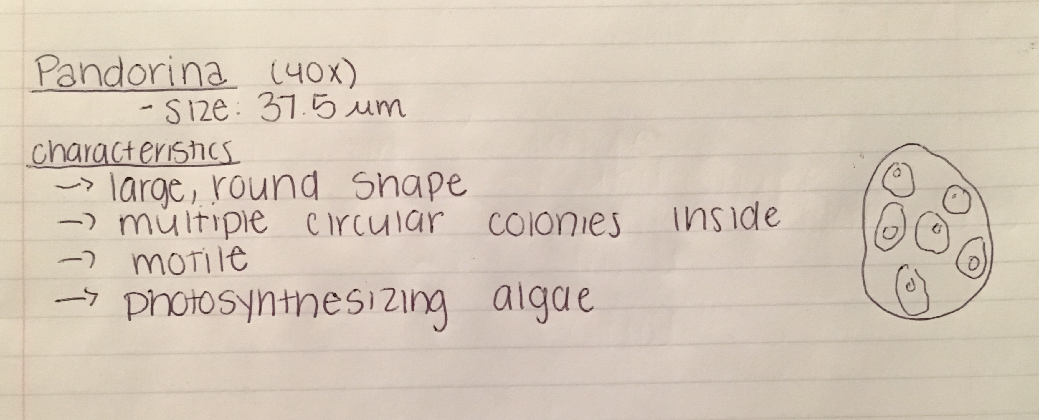
Data and Observations: When we brought back our hay infusion we noted that it had no noticeable scent. It was a brown and green murky color and had tan clusters of film present on the top. We obtained three organisms from the top, chilomonas (22um), colpidium(51 um), and paramecium aurelia (156um). All three of these organisms were motile, and protozoa - thus making them non-photosynthetic.
These images are of the organisms respectively :
File:IMG 0891.jpeg
File:IMG 0890.jpeg

From the bottom sample of our hay infusion we found peranema (45 um), arcella (63um), and paramecium bursaria (156um). These organism were also all motile, non-photosynthesizing protozoa.
These are images of the organisms respectively:
File:IMG 5072.jpeg


Conclusions and Future Directions: This weeks lab really emphasized the scale of organisms, and illustrated the wide spectrum of life present just on our own campus. For instance, and organism such a paramecium meets all the needs of life: energy, cells, replication, information, and evolution. The paramecium feeds on other microorganisms for energy, is unicellular, reproduces asexually through binary fission, has its own genome of information, and has evolved over time to better fit its environments (like its movement methods). If our hay infusion "grew" for another two months I suspect that there would be a developing shortage of food and space within the small jar and it would result in perhaps the dying off of these organisms (assuming that no additional nutrients were added).
ST
1.27.15
Good first entry. Need to include more detail for example there is no mention of the Hay Infusion set-up. Pictures would add information. The transect was 20Ft by 20Ft, not meters, there is a typo in the manual.
SK
1/26/15
Lab 1: Biological Life at AU
Purpose: The purpose of this lab is to observe and analyze the ecological interactions occurring in our given “niches” represented by a 20 x 20 meter transect of land somewhere on campus. We will explore the sheer amount of biological diversity present just within American University’s campus alone through weekly visits to our given niches.
Materials and Methods: Each group was assigned a transect of land that is 20 x 20 meters large and we are responsible for observing and analyzing the ecology of the environment and recording data. The transect of land is the AU student run community garden behind Leonard Hall next to the campus tennis courts. We are responsible for weekly observations of our plot and data recording.
Data and Observations: We observed the agricultural nature of the transect which included an irrigation system(hoses running through each separate garden box), presumably fertilized soil(only in the garden boxes), and of courseseparate wooden garden boxes for each different plant. The plants present in our transect of the garden are brussel sprouts, lettuce, kale, and cucumbers. In addition to the more uniformed boxes, there is an area of stray leaves, mulch, and hay on the edge of our transect.
Biotic features of our transect included things such as: pests, rodents, birds, weeds, vegetables, earthworms, etc. While abiotic features included: air, water, soil, mulch, etc. One prominent factor that will determine the state of our transect is human interaction because it is a garden that depends on irrigation and human maintenance.
Conclusions and Future Directions: I think it will be interesting to observe the qualities in our transect in contrast to the more naturally occurring transects that students have - as our transect is so influenced by humans. I also am interested to see the interactions between the vegetables and the attraction of pests to the garden.
1/22/15
Test


