User:Student 66/Notebook/Biology 210 at AU: Difference between revisions
No edit summary |
No edit summary |
||
| Line 2: | Line 2: | ||
BIO-210-007 | BIO-210-007 | ||
2/4/2016 | 2/4/2016 | ||
---- | |||
'''Week 2: Hay Infusion''' | '''Week 2: Hay Infusion''' | ||
| Line 33: | Line 36: | ||
11. Repeat this process on the +tet plate labeled 10^-3. | 11. Repeat this process on the +tet plate labeled 10^-3. | ||
12. Repeat this procedure with the 10^-4 | 12. Repeat this procedure with the 10^-4 dilution on the 10^-5 plates, 10^-6 dilution on the 10^-7 plates and the 10^-8 dilution on the 10^-9 plates. | ||
13. Set the agar plates aside (side up) onto a rack. Leave them there at room temperature for a week. | 13. Set the agar plates aside (side up) onto a rack. Leave them there at room temperature for a week. | ||
| Line 43: | Line 46: | ||
[[Image:JarBAM.jpg]] | [[Image:JarBAM.jpg]] | ||
Figure 1: | Figure 1: Transect #3 Jar | ||
[[Image: | [[Image:JarBAM3.jpg]] | ||
Figure 2: Transect #3 Jar from above | |||
1. The smell was pungent, like sewer. Almost a mold-like smell. Murky in appearance. The water was a light brown/green. | 1. The smell was pungent, like sewer. Almost a mold-like smell. Murky in appearance. The water was a light brown/green. | ||
2. There was life on top of the liquid, as seen by the mold film on the surface of the water. | 2. There was life on top of the liquid, as seen by the mold film on the surface of the water. | ||
[[Image:JarBAM2.jpg]] | |||
''Niches of our jar:''' | ''Niches of our jar:''' | ||
[[Image:JarBAMniches.jpg]] | [[Image:JarBAMniches.jpg]] | ||
Figure 3: Jar Niches - Three distinct niches including: top, middle, bottom | |||
4. Protists and algae present | 4. Protists and algae present | ||
| Line 71: | Line 78: | ||
[[Image:MR1.jpg]] | [[Image:MR1.jpg]] | ||
Figure 4: Gonium found top niche. | |||
The following organism was found in the top layer of our jar. Observed was the gonium (colony up to 90 um). | The following organism was found in the top layer of our jar. Observed was the gonium (colony up to 90 um). | ||
| Line 77: | Line 85: | ||
[[Image:MR2.jpg]] | [[Image:MR2.jpg]] | ||
Figure 5: Volvox found middle niche. | |||
The following organism was found in the middle layer of our jar. Observed was the volvox (350-500 um). | The following organism was found in the middle layer of our jar. Observed was the volvox (350-500 um). | ||
| Line 83: | Line 93: | ||
[[Image:MR3.jpg]] | [[Image:MR3.jpg]] | ||
Figure 6: Didinium cyst found on bottom layer. | |||
The following organism was found in the bottom layer of our jar. Observed was the didinium cyst. | The following organism was found in the bottom layer of our jar. Observed was the didinium cyst. | ||
| Line 94: | Line 105: | ||
[[Image:SerialDBAM.jpg]] | [[Image:SerialDBAM.jpg]] | ||
Figure | Figure 7: Serial Dilution Process | ||
'''Conclusion''' | '''Conclusion''' | ||
Our hypothesis appeared | Our hypothesis appeared to be correct. There were different protists and algae found in the different niches. | ||
Revision as of 12:48, 4 February 2016
Madeline Rohrbacher BIO-210-007 2/4/2016
Week 2: Hay Infusion
Introduction The purpose of the following experiment to observe the unicellular eukaryotic organisms growing on the top, middle, and bottom of Transect #3's Hay Infusion Culture. We used a dichotomous key to identify the protists and algae found in Transect #3. Our hypothesis would be that the different niches (top, middle, bottom) would have different protists and algae due to the different abiotic and biotic components in each layer.
Materials and Methods
1. Observe the Hay Infusion Culture. Make any notes on the appearance, smell, and color.
2. Identify the different layers of the Hay Infusion Culture and describe each.
3. Observe a sample from each niche.
4. Measure each organism and identify each using a dichotomous key.
5. Mix the Hay Infusion Culture jar by swirling it around with the lid on.
6. Collect and label four tubes of 10mLs sterile broth with 10^-2, 10^-4,10^-6,10^-8. Also collect a micropippetor and set is at 100micro Liters.
7. Collect four nutrient agar and four nutrient agar plus tetracycline plates. Label the plates with tetracycline with "tet" - always on the side of the plate.
8. Add 100 micro Liters from the culture to the 10mLs of broth in the tube labeled 10^-2. Swirl the inoculated tube throughly.
9. Repeat this process twice more to make the 10^-6 and 10^-8 dilutions.
10. Pipette 100 micro Liters from the 10^-2 tube onto the nutrient agar plate labeled 10^-3.
11. Repeat this process on the +tet plate labeled 10^-3.
12. Repeat this procedure with the 10^-4 dilution on the 10^-5 plates, 10^-6 dilution on the 10^-7 plates and the 10^-8 dilution on the 10^-9 plates.
13. Set the agar plates aside (side up) onto a rack. Leave them there at room temperature for a week.
Results
Observations of Hay Infusion Culture
Figure 1: Transect #3 Jar
Figure 2: Transect #3 Jar from above
1. The smell was pungent, like sewer. Almost a mold-like smell. Murky in appearance. The water was a light brown/green.
2. There was life on top of the liquid, as seen by the mold film on the surface of the water.
Niches of our jar:'
Figure 3: Jar Niches - Three distinct niches including: top, middle, bottom
4. Protists and algae present
On the top layer: Gonium, pandorina, colpidium, volvox
Middle layer: Volvox, paramecium bursaria, spirostomum, eugirna
Bottom layer: Blepharisma, didinium cyst, chilomonas sp,, spirostomum, pandorina colony
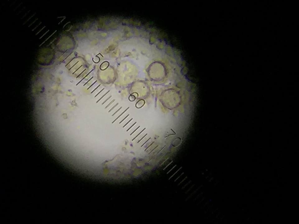
 Figure 4: Gonium found top niche.
The following organism was found in the top layer of our jar. Observed was the gonium (colony up to 90 um).
 Figure 5: Volvox found middle niche.
The following organism was found in the middle layer of our jar. Observed was the volvox (350-500 um).
 Figure 6: Didinium cyst found on bottom layer.
The following organism was found in the bottom layer of our jar. Observed was the didinium cyst.
6. None of the following above were motile.
7. If the Hay Infusion Culture "grew" for another two months, the
Serial Dilution:
Figure 7: Serial Dilution Process
Conclusion
Our hypothesis appeared to be correct. There were different protists and algae found in the different niches.
Transect #3
Week 1: Transect Observation
Location:
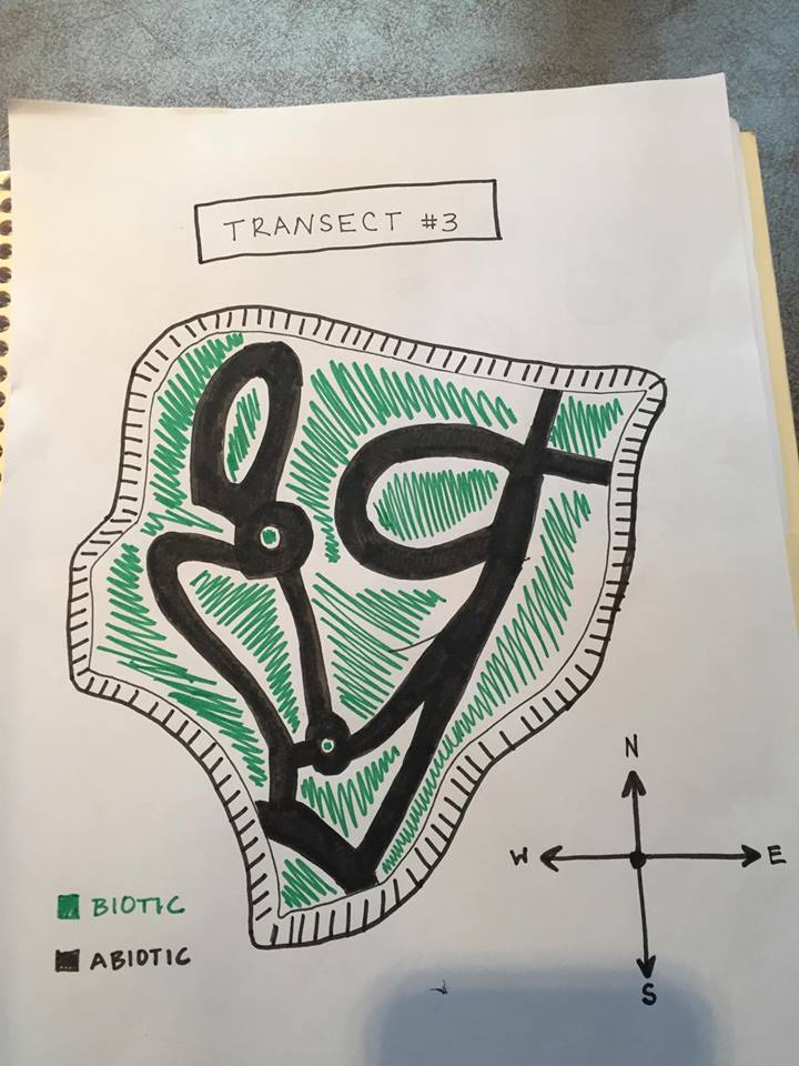
Aerial view of Transect showing Abiotic and Biotic areas:
Transect #3 sits between Bender Arena and Hughes Residence Hall. The transect lies just above the amphitheater and before the road. The area consists of a few benches, light poles, garbage can, and a concrete pathways winding about the garden. The ground is relatively flat only to be interrupted with a staircase. The soil is dark with mulch and dead leaves on top of it. There are a variety of trees and plants assorted throughout the transect as well.
Abiotic features: 1. Metal benches 2. Mulch 3. Rocks 4. Dirt 5. Lamp poles
Biotic features: 1.Leafy plants 2. Leaves 3. Weeds 4. Tall trees 5. Small insects
Madeline_B._Rohrbacher


