User:Suzanne Shaffer/Notebook/Biology 210 at AU: Difference between revisions
No edit summary |
No edit summary |
||
| Line 25: | Line 25: | ||
Gram Stain Observations: | Gram Stain Observations: | ||
The Gram staining is the use of crystal violet to see if the cell walls have a thick layer with peptidoglycan. If the cell is darkened only on the outside it means that this thickened membrane has taken up this dye into its cell membrane and is positive. If the cell appears all dark, this means that there is a phospholipid layer to the membrane which prevents this reuptake of crystal violet. Upon looking at our three samples it was observed that 10^-5 was gram positive because the hundreds cocci cells only had a darkened outside of the circle with clear on the inside. For sample 10^-7 and 10-4, the rods and cocci both appeared all dark indicating that they are gram negative. | The Gram staining is the use of crystal violet to see if the cell walls have a thick layer with peptidoglycan. If the cell is darkened only on the outside it means that this thickened membrane has taken up this dye into its cell membrane and is positive. If the cell appears all dark, this means that there is a phospholipid layer to the membrane which prevents this reuptake of crystal violet. Upon looking at our three samples it was observed that 10^-5 was gram positive because the hundreds cocci cells only had a darkened outside of the circle with clear on the inside. For sample 10^-7 and 10-4, the rods and cocci both appeared all dark indicating that they are gram negative. | ||
[[Image:Staining 1.JPG]] | |||
Revision as of 20:47, 16 February 2014
2/16/2014 Question/Objective: Looking at hay infusion bacteria, which was cultured on nutrient and tetracycline plates note characteristics of the different bacteria. Selecting three colonies perform wet mount for cell observation, gram straining and PCR. Steps: 1.) Check on Hay Infusion and make observations on the changes that may have occurred over the week. 2.) Obtain the 7 cultures that were plated last lab and Complete Data Table 1 100-fold Serial Dilutions Results. Counting the number of colonies that have grown on the nutrient and tetracycline plates. 3.) Pick three of these plates at least one tetracycline and fill in data on Data Table 2. 4.) Note the size and characteristics of one colony on each plate. 5.) Wet mount a tiny bit of each of these three colonies mixing it with a drop of oil then placing a cover slip on top. Observe the sample under the microscope at 10x 40x and 100x. Note the cell descriptions of the cells including their shapes and arrangement. 6.) Perform gram staining. Take another tiny sample three bacteria colonies and again on three separate slides. The sample needs to be dried, so no water or cover slip is added, each sample is passed through a flame about 8-9 times or until appearing dry. Then each sample is washed with 1 minute of crystal violet, rinsed with bottled water, Gram’s iodine mordant for 1 minute, rinse with water, 95% alcohol for decolorizing for 10-20 seconds, washed with safranin stain for 20-30 seconds. Finally the samples are rinsed with water again and carefully dried before examination. Observe to identify all three sample as gram positive or negative. 7.) Perform PCR on a sample of the hay infusion for examination in next class. Mixing tiny amount of hay infusion sample with 100 microliters of water. Place this on a heat block for 10 minutes. After using the centrifuge, the sample is spin for 1 minute. With the DNA in the supernatant, pipette out 2 microliters of the DNA and mix with 23 microliters of the reverse primers. Place initials on the tiny tube and place into the PCR machine.
Observations: Hay Infusion observations: Upon analysis of the hay infusion this week it was noted that there were some changes to the environment. There was no film of algae at the top of the water as there previously was. Also, the potent smell that it had last week has decreased significantly. There was notable decrease in the water and the water was actually clearer with increased sediment on the bottom. These changes are due to the lack of nutrients that there is after one week. There is a disappearance of certain organisms due to the lack of energy required to sustain life, thus changes such as the smell decrease as well and the water being more clear.
Serial Dilution Results: Table 1 100 Fold Serial Dilution shows the colonies counted and colonies per milliliter results from the 4 nutrient and 3 tetracycline treated plates. Its was obvious to observe that the greater the dilution, for both the nutrient and tetracycline treated plates, the less colonies that grew on the plate. It was also noticed that the tetracycline treated plates showed significantly decrease in colony growths but also were larger in size when comparing to the nutrient plates. This tells us that the nutrient plates, had more colonies and they were smaller in size because they were growing on top of one another. Being that tetracycline is an antibiotic, the only growths on these three plates were those, which were antibiotic resistant to tetracycline. Tetracycline is a board spectrum antibiotic like many used for both gram-positive and gram-negative bacteria. The antibiotic affects the protein synthesis within the bacteria. So there were few growths of bacteria on these plates and no fungus present. It is known that there are approximately 29 different genes that are unaffected by tetracycline (Chopera & Roberts, 2001).
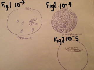
Bacteria Cell Morphology Observations: The samples that were chosen for further observation were plates 10^-5 (nutrient), 10^-7 (nutrient) and 10^-4 (tet). First the samples were placed onto a slide with oil and observed at 100x. It was obvious that plate 10^-7 (Figure 1) had the rod shape and was classified as a diplobacilli. Looking at 10^-4 (figure 2) there were hundreds of cocci also noted and was classified as staphylococcus. The last one observed, 10^-5 (figure 3), was unable to be classified under the microscope. There was a 3D appearance to this sample and even after getting another sample and staining, it was still not visible under the microscope. The professor was notified and stated that we might be able to visualize the shape after staining.
Gram Stain Observations: The Gram staining is the use of crystal violet to see if the cell walls have a thick layer with peptidoglycan. If the cell is darkened only on the outside it means that this thickened membrane has taken up this dye into its cell membrane and is positive. If the cell appears all dark, this means that there is a phospholipid layer to the membrane which prevents this reuptake of crystal violet. Upon looking at our three samples it was observed that 10^-5 was gram positive because the hundreds cocci cells only had a darkened outside of the circle with clear on the inside. For sample 10^-7 and 10-4, the rods and cocci both appeared all dark indicating that they are gram negative.
2/6/14 lab 1 notes
Great job! Start working on building a map of your transect to detail your land and where your samples are taken from. We will talk about this more Wednesday. Great work!
AP
January 22, 2014 Day 2: Hay Infusion Culture Observations: Question/Objective: With the understanding of a dichotomous key be able to identify protists from ones niche.
Steps: 1) Looking at the Hay Infusion write down the changes that have occurred over the last week. 2) Take three samples from the hay infusion and put them on a slide with a coverslip 3) Using the dichotomous key identify 6 protist that were observed on the slide. 4) Draw pictures of these organism that were found and obtain measurements and characteristics. 5) Prepare serial dilutions: Gather 4 test tubes labeling even them 2-8. Place 10 ml in each tube. Using a 100 microliter pipette put 100 microliters of hay infusion sample into the 1st test tube. Mix this solution then take out 100 microliters and place this into the 2nd test tube. Repeat until test tube 4. 6) Obtaining 4 nutrient agar plates from the instructor label each –3, -5, -7,-9. Then obtain 3 nutrient agar plates with tetracycline and label then –2,-4,-6. 7) Starting with test tube 8 pipette 100 microliters of sample onto the plate that is labeled –9 and spread the sample evenly around the plate, continue with tube 6 and plate –7 and so forth. 8) The same procedure for the plates with the tetracycline starting with plate –6 and sample from test tube 6. Continue for –4 from test tube 4 and –2 from test tube 2.
Observations/Data:
Hay Infusion Observations: Looking at the ecosystem that was created last class, it is easy to see that there have been a lot of changes in one week. Firstly, the smell, which was a dirt/soil smell, has changed to a potent/harsh moldy smell. Upon initial assessment of the jar, the water was much darker dirt brown with increased black sediment on the bottom of the jar. There was a clear film that was visible across the top of the water. There was new growth on some of the pine needles and leaves within the liquid. It is predicted that in two months there will be even more growth to the hay infusion. The hay infusion will continues to have growths of bacteria, protist and algae as the weeks go on as long as the temperature is held the same, adequate sunlight, food/energy and oxygen. The growth will plateau if any of these conditions change in the next two months. Inadequate temperature, sunlight, food/energy and oxygen would result in the selective pressures, which are pressures that cause the reproduction to decrease in the environment.
Sample Selection: When looking at the ecosystem with the group it was decided where the three samples needed to come from. The first was from the top, when taking this sample it was very difficult to penetrate the film that was on the top of the water. The second sample was from the middle around the needles and the third was from the bottom where there was a lot of darker sediment. These different areas should theoretically yield different types of species. One would expect that species that are living near other plants matter rely on those plants for energy. On the other hand, species on the top of the water are able to produce energy from other forms such as photosynthesis. Figure one: Hay Infusion Culture Observations:
In order for an organism to be considered alive they have to meet certain requirements. They must be able to obtain and utilize energy, be made up of cells, process information through DNA and RNA, have the ability to reproduce and of course come from other cells/being able to evolve (Freeman, 2010). Looking at the amoeba that was found in group 5’s hay infusion it is evident that it is a living thing and has all these qualities of living things. Amoebas obtain food by engulfing it (decaying vegetation), as it moves in a slow creeping way (Freeman, 2010). It is a protist that has a nucleus with genetic information and can be unicellular or multicellular. They are able to reproduce through the process of meiosis to form spores (Freeman, 2010). Amoebas have evolved from and there is many different types exemplifying that the species has been capable of evolution.
Figure 2: Serial Dilutions:
Future Plans: With the hay infusion solution plated on the agar, the bacterial will grow on the plates and will be examined next week.
Citation: Freeman, Scott. 2002. Biological Science. Prentice Hall: New Jersey. (2, 569-570).
January 22, 2014 Lab notebook and username entered successfully. -SMS
great work! MB
January 16, 2014 Day 1: Visit Transect to Study and Obtain Sample for Hay Infusion
Question/Objective: Given a 20x20 transect, analyze the land on the American University Campus and describe the general characteristics of the area including the biotic and abiotic components.
Steps: 1- TA walked group 5 to the transect area by the Church on Massachusetts Ave. 2- Having area cleared by Ben, a sample was collected from the transect. The sample included about 50% of the dirt/soil and the other the leaves/needles/vines and ivy. It was collected this sample in a 50ml conical tube using metal shovels. 3- Then 11.99g of the sample was measured out and placed into a labeled plastic jar, which had 500ml of water in it. 4- After adding 0.1g of dried milk to the sample in the water, it was gently mixed by swirling for approximately 10 seconds. 5- This transect was placed in the back of the room in the lab.
Observations: General: In the 20x20 section that group 5 was given was in a very dark shady area. The darkness was from the church and the trees in the area. Trees: There are three large trees in the area. The first one was a holly tree with scant amount of red berries on it and was healthy looking/damp from the previous days rainstorm. The second tree was the tallest which appeared to be dead. There was an area where the bark was missing; there was no leaves or evidence of recent leaves on this tree. The last tree is a pine tree with lots of pine needles still attached and healthy looking. This tree was also damp from the rain. Ground: On the ground there was lots of ivy that covered nearly the whole 20x20 area that was provided. There were lots of leaves that were from the trees, including the spiky brownish lifeless pine needles, maple leaves, acorns, and holly leaves. There were lots of small branches of each of the trees noted in this area as well. Under the ivy there was soil, mulch and vines from the trees and the ivy. There were no animals/insects noted in the area that were observed during this initial visit to the intersect. Abiotic: water, soil, air, and minerals in the dirt Biotic: trees, fungi on the tree trunks, leaves, ivy
Future Plans: The hay infusion, which was collected, will stay in the lab for another week. Observations will be documented after one week and prokaryotes will be examined from two locations under the microscope.-SMS


