Biomod/2014/NTU/Review
<html> <head> <title></title> <link href="http://openwetware.org/index.php?title=Biomod/2014/NTU/Templates/css/bootstrap.css&action=raw&ctype=text/css" rel='stylesheet' type='text/css' /> <link href="http://openwetware.org/index.php?title=Biomod/2014/NTU/Templates/css/animate.css&action=raw&ctype=text/css" rel="stylesheet" type="text/css" media="all"> <link href="http://openwetware.org/index.php?title=Biomod/2014/NTU/Templates/css/style.css&action=raw&ctype=text/css" rel='stylesheet' type='text/css' /> <script type="text/javascript" src="http://openwetware.org/index.php?title=Biomod/2014/NTU/Templates/js/jquery.min.js&action=raw&ctype=text/js"></script> <script type="text/javascript" src="http://openwetware.org/index.php?title=Biomod/2014/NTU/Templates/js/slidejs&action=raw&ctype=text/js"></script> <script src="http://openwetware.org/index.php?title=Biomod/2014/NTU/Templates/js/slidediscussion&action=raw&ctype=text/js"></script> <script> $(function () { $("style[media*='screen']").remove(); $("link[href*='favicon']").remove(); //fix heading var h1 = $(".firstHeading").text().split("/"); $(".firstHeading").text(h1[h1.length-1]); $("tr:odd").addClass("odd"); }); function slideload(){ $('#fadein').fadeIn(6000); }
                var urlstr=location.hash;
                if(urlstr=="#1")
                {
                                $("html body").animate({
                scrollTop: 2000
               },1000);
                document.location.href.replace(location.hash , "" );
               };
                if(urlstr=="#2")
                {
                                $("html body").animate({
                scrollTop: 3100
               },1000);
                document.location.href.replace(location.hash , "" );
               };

                if(urlstr=="#3")
                {
                                $("html body").animate({
                scrollTop: 2600
               },1000);
               document.location.href.replace(location.hash , "" );
               };
</script> <style>
.body {
 width:100%;
 color: #CCCCCC ;
 max-width: 1280px;
 min-width: 0 ;
 padding-top: 55px;
 margin-left: auto;
 margin-right: auto;
}
- column-content #content {
   padding: 0em;
   margin: 0;
}
- content{
 border: 0px;
 float:right;
 padding: 0em;
 width:100%;
}
- contentSub, #search-controls, .firstHeading, #footer-box, #catlinks, #p-logo, #toctitle ,#top-section ,#column-one ,#footer, #siteSub,#jump-to-nav,.printfooter,.visualClear
{
display:none;
}
- globalWrapper {
 background-color:#fff;
 font-size:100%;
 padding-bottom: 0px;
}
.start:hover{ background-color: #CE7000; transition: background-color 0.5s linear; } </style> <link href="http://openwetware.org/index.php?title=Biomod/2014/NTU/Templates/css/slidecss&action=raw&ctype=text/css" rel='stylesheet' type='text/css' /> </head> <body onload="slideload()" style="background-color:#">
- <a href="http://openwetware.org/wiki/Biomod/2014/NTU"><img src="http://openwetware.org/images/4/4d/Logo1.png" style="left:-50px;"></a>
- <a href="javascript: void(0)">Project</a>
- <a href="http://openwetware.org/wiki/Biomod/2014/NTU/Idea">Idea</a>
- <a href="http://openwetware.org/wiki/Biomod/2014/NTU/Design">Design</a>
- <a href="http://openwetware.org/wiki/Biomod/2014/NTU/Origami">Origami</a>
- <a href="javascript: void(0)">Experiment</a>
- <a href="http://openwetware.org/wiki/Biomod/2014/NTU/Method">Method</a>
- <a href="http://openwetware.org/wiki/Biomod/2014/NTU/Result">Result</a>
- <a href="javascript: void(0)">Discussion</a>
- <a href="#">Review</a>
- <a href="http://openwetware.org/wiki/Biomod/2014/NTU/Future">Future</a>
- <a href="javascript: void(0)">Supplement</a>
- <a href="http://openwetware.org/wiki/Biomod/2014/NTU/Protocol">Protocol</a>
- <a href="http://openwetware.org/wiki/Biomod/2014/NTU/Material">Material</a>
- <a href="http://openwetware.org/wiki/Biomod/2014/NTU/Equipment">Equipment</a>
- <a href="http://openwetware.org/wiki/Biomod/2014/NTU/Member">Member</a>
- <a href="http://openwetware.org/wiki/Biomod/2014/NTU/Acknowledge">Acknowledge</a>
Purification
According to our speculation, the reason behind our failure is that we did not succeed in purifying our DNA origami product. Purification is one of the most important steps, and the excessive impurities that remained may have interrupted with our subsequent experiments.
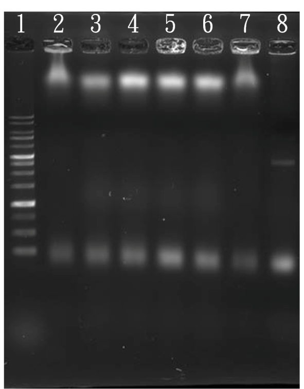
For the purification, we used the Amicon Ultra-0.5 Centrifugal Filters, which is supposed to filter out any molecules smaller than 100 kilodalton. The result is shown in the picture below.
<img src="http://openwetware.org/images/3/31/Discussion1.png" style="position:relative; margin-left:150px;"/>
From left to right:
1. 1kb marker
2. Monomer without purification
3. Monomer purified once
4. Monomer purified twice
5. Monomer purified three times
As we can see, there is no significant difference between the jellyfish before and after purification. The primers that we want to filter out are still present, even after 3 cycles of purification. We have struggled with the problem of purification for a long time. Due to the limited time we had, we were unable to successfully complete the purification procedure.
Such disappointing result prevented us from doing some of our experiment, which were components 7(to test whether protection strand works) and 8(to test whether complementary strand of protection strand can be removed.) Unpurified sample would contain many suspended protect strand in the solvent, this condition made the design of its complementary strand useless.
To continue our project, some steps must fine tune a little. For example, we actually mixed the outside aptamer and its complementary strand together before we added the monomer. So did our inner aptamer and the poly C or G strands. That process lead to better production rate of dimer and aggregation effect.
TEM
Although we can see the monomers and dimers under TEM, we are unable to quantify the products. It is therefore difficult to tabulate specific results such as monomer production and dimer association rate. In addition, we often saw giant black geometries and small white circles under TEM, which could be attributed to sample contamination. The unclear membrane surface may contain these contaminations, which may interfere with our products. However, we did not find significant defects in TEM pictures of our sample.
Poly C-G aggregation and virus capture
<img src="http://openwetware.org/images/d/d4/Discussion2.png" style="position:relative; margin-left:150px;"/>
Ideally, there should be tailing in the 4th well, in which jellyfish should aggregate because of poly C-poly G hybridization (Watson-Crick hybridization). However, we can't see anything at the same place of control. This failure could be due to the insufficient amount of DNA for ETBR staining.
<img src="http://openwetware.org/images/a/a4/Discussion3.png" style="position:relative; margin-left:150px; top:-80px;"/>
The same situation happened in this experiment, too. Dimers reacted with virus from well 3 to 6, and the tailing phenomenon is invisible. We also thought that the insufficient amount of DNA for ETBR staining caused the failure.
The upper band was the GFP, and apparently it is slightly slower in the wells 2 and 7. Considering the darker primers below, we believed that this phenomenon resulted in the interaction between positive charge of allantoic protein and GFP. Furthermore, the negative charge of our dimer interacted with the allantoic protein, so the band disappeared.
For basic information of above two figures, please check our results.
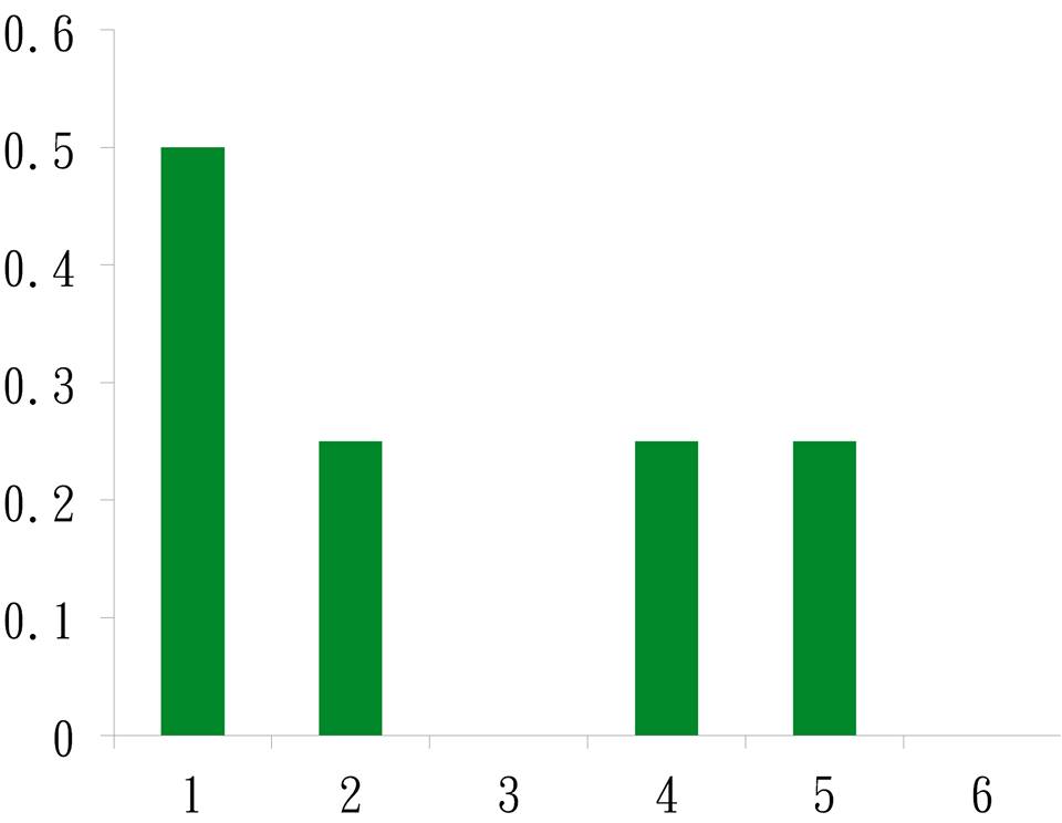
Hemagglutination assay
<img src="http://openwetware.org/images/c/c5/Discussion4.png" style="position:relative; margin-left:150px;"/>
The Y-axis represents virus activity by HA test. Number 2,4 and 5 represent dimers reacted with virus. Number 1 holds the virus, and number 3 and 6 are negative control mixed with dimer and unpurified allantoic proteins. Our product seems to reduce the activity of virus proteins.
<img src="http://openwetware.org/images/1/17/Review.jpg" style="position:relative; left:150px;">
However, to ensure that all aptamer binding sites in the monomer can be occupied by either a lock or an inner aptamer successfully, we set the concentration of aptamer: monomer as 3:1. When the “monomer +aptamer” and “virus” samples are mixed together, the excessive free aptamers will bind to the H5 proteins. Due to the incomplete purified nature of our sample, it is possible that part of the inactivation might be a result of suspended aptamers.
The upper band was the GFP, and apparently it is slightly slower in the wells 2 and 7. Considering the darker primers below, we believed that this phenomenon resulted in the interaction between positive charge of allantoic protein and GFP. Furthermore, the negative charge of our dimer interacted with the allantoic protein, so the band disappeared.
For basic information of above two figures, please check our results.