Kubke Lab:Research/CND/Records2010-2011Summer/RC015
| Cranial Nerve Development | Experiment |
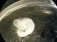
Embryo details
Species: Gallus gallus domesticus
Embryo Name: RC015

Embryo stage: ST20 + Confirmed by Fabiana (supervisor).
Staging description: Staged on the 20th Jan 2011 10:00am, see Notebook Entry
- Tail bud points forwards
- Leg and wing buds roughly the same size (indicative of an embryo ST20-ST21)
- Faint pigmentation present in the eye (pigmentation begins to develop at ST20)
- Maxillary visceral arch is distinctly larger than the Mandibullary which extends to midline of eye (indicating ST20-ST21)
- 2nd Visceral arch distinct. (Indicating ST20-ST21).
Fixation: PFA
Cryoprotection: None.
Material label and storage: The sectioned, mounted, stained and coverslipped (in DPX) embryo is labeled RC015 and stored in a slide folder in the lab.
Experiment details
Objective:To complete a serial coronal sectioning of the embryo rostral to the wingbud prior to staining with Cresyl Violet and coverslipping for histological and cytological analysis of the stained sections.
Procedure:
20th Jan 2011, 10:15am (see Notebook entry)
- The embryo RC015 was staged according to the Hamburger and Hamilton (1951) staging system.
- 11:30am Using dissection scissors and under a dissection microscope I cut the embryo immediately rostral to the wing bud
- The cryostat was set to OT: -19°C, CT: -19°C and allowed to equilibrate to the new temperature (it was previously set to OT:-17°C, CT: -17°C.
- The head region of the embryo was then gently replaced into a vial filled with PFA using forceps.
- A small plastic mould was half filled with OCT and then, using a blade, the embryo was very carefully laid on top of the OCT from a Petri dish containing PFA.
- Using forceps the embryo was oriented such that the hindbrain ran parallel to the sides of the mould so that coronal sections could be made
- Bubbles in the OCT were removed with the forceps.
- 11:55am Incubated the block at -19°C in the cryostat chamber.
- 12:15pm The block was oriented 90° to a chuck and stuck on by freezing OCT between the two contacting surfaces.
- OCT was built up either side of the block inside the cryostat chamber.
- The chuck was inserted into the metallic chuck holder and incubated at -19°C for one hour.
- 1:15pm Began trimming the block but decided once I had trimmed to the embryo that it was not centered properly and so more OCT was built up on the side lacking OCT to centre the tissue in the OCT mould.
- The assembly was incubated in the metallic chuck holder for 30 minutes prior to cutting to allow it to harden.
- Serial sections of the specimen were cut and mounted onto microscope slides and dried overnight in a fume hood.
- Slides 1,3,5,7,9,10,11 & 15 were cut by Reuben and Slides 2,4,6,8,12,13,14 were cut by Malisha.
21st Jan 2011, 9:30am see Notebook entry
- A Cresyl Violet stain was then done by Reuben on the tissue sections prior to coverslipping and storing the slides in the folder labeled RC015.
Cryostat Sectioning Cryostat settings (for a more detailed protocol visit Kubke_Lab:Cryomicrotomy)
| Cryostat | Leica – CM3050S |
| Knife | MX35 Premier +, 34 degrees, 80mm Thermo Scientific |
| Day Cut | 20th Jan 2011, 1:15pm (see Notebook entry) |
| Knife Angle | 1.5° |
| Chamber Temp | -19°C |
| Object Temp | -19°C |
| Glass Slides | Gelatin-subbed Original Menzel-Glaser microscope slides with cut edges and frosted Ends. Slides were subbed using the Cold gelatin subbing protocol including the pre-wash procedure. |
| Plane of section | |
| Number of slides | 15, 204 sections |
| Observations | Sections 24,131, 195 and 197 were lost during mounting. One section lifted off its slide and was found in the mounting Xylene. Further investigation to determine which section this was is needed. Section 118 was badly damaged but still mounted. Sections from slide 4 are noticeably damaged. |
(Include in your observations, eg ,were the sections serial, was any section lost, was quality assessed, etc)
Cresyl Violet staining
For more informattion see Kubke_Lab:Nissl_Stain_Protocol
| Date | ||
| Defatting and rehydration step | ||
| Solution | Time | Comments |
| Water | 1 Dip | |
| 70% alcohol | 4 mins | |
| 95% alcohol | 4mins | |
| 100% alcohol 1 | 4mins | |
| 100% alcohol 2 | 4mins | |
| Xylene 1 | 5mins | |
| Xylene 2 | 5mins | |
| Xylene 3 | 5mins | |
| Xylene 2 | 2mins | |
| Xylene 1 | 2mins | |
| 100% alcohol 2 | 2mins | |
| 100% alcohol 1 | 2mins | |
| 95% alcohol | 2mins | |
| 70% alcohol | 2mins | |
| Water | 2 dips | |
| Staining and differentiation step | ||
| Solution | Time | Comments |
| Water | 2dips | |
| Cresyl Violet | 5mins | |
| 50% alcohol | 2mins | |
| 70% alcohol acetic acid | 0 | Did not prepare the solution in time for the experiment. |
| 95% alcohol | 2mins | |
| 100% alcohol 1 | 2mins | |
| 100% alcohol 2 | 2mins | |
| Xylene 1 | 2mins | |
| Xylene 2 | 2mins | |
| Xylene 3 | 2-25minutes | See table below for times spent in Xylene prior to coverslipping. |
| Coverslip | Ind. | |
- (Note: Please incorporate the information on the table below with the table above--MF Kubke 05:12, 3 February 2011 (EST))
| Slide Number | Time spent in Xylene (mins) |
|---|---|
| 14 | 2 |
| 13 | 3 |
| 12 | 5 |
| 8 | 7 |
| 16 | 8 |
| 6 | 9 |
| 4 | 10 |
| 2 | 12 |
| 15 | 13 |
| 9 | 15 |
| 11 | 16 |
| 7 | 17 |
| 10 | 18 |
| 3 | 20 |
| 5 | 22 |
| 1 | 25 |
Comments:


