Biomod/2014/NTU/Design
<html> <head> <title></title> <link href="http://openwetware.org/index.php?title=Biomod/2014/NTU/Templates/css/bootstrap.css&action=raw&ctype=text/css" rel='stylesheet' type='text/css' /> <link href="http://openwetware.org/index.php?title=Biomod/2014/NTU/Templates/css/animate.css&action=raw&ctype=text/css" rel="stylesheet" type="text/css" media="all"> <link href="http://openwetware.org/index.php?title=Biomod/2014/NTU/Templates/css/style.css&action=raw&ctype=text/css" rel='stylesheet' type='text/css' /> <script type="text/javascript" src="http://openwetware.org/index.php?title=Biomod/2014/NTU/Templates/js/jquery.min.js&action=raw&ctype=text/js"></script> <script type="text/javascript" src="http://openwetware.org/index.php?title=Biomod/2014/NTU/Templates/js/slidejs&action=raw&ctype=text/js"></script> <script src="http://openwetware.org/index.php?title=Biomod/2014/NTU/Templates/js/slideproject&action=raw&ctype=text/js"></script> <script> $(function () { $("style[media*='screen']").remove(); $("link[href*='favicon']").remove(); //fix heading var h1 = $(".firstHeading").text().split("/"); $(".firstHeading").text(h1[h1.length-1]); $("tr:odd").addClass("odd"); }); function slideload(){ $('#fadein').fadeIn(6000); } </script> <style>
.body {
 width:100%;
 color: #CCCCCC ;
 max-width: 1280px;
 min-width: 0 ;
 padding-top: 55px;
 margin-left: auto;
 margin-right: auto;
}
- column-content #content {
   padding: 0em;
   margin: 0;
}
- content{
 border: 0px;
 float:right;
 padding: 0em;
 width:100%;
}
- contentSub, #search-controls, .firstHeading, #footer-box, #catlinks, #p-logo, #toctitle ,#top-section ,#column-one ,#footer, #siteSub,#jump-to-nav,.printfooter,.visualClear
{
display:none;
}
- globalWrapper {
 background-color:#fff;
 font-size:100%;
 padding-bottom: 0px;
}
.start:hover{ background-color: #225E94; transition: background-color 0.5s linear; } </style> <link href="http://openwetware.org/index.php?title=Biomod/2014/NTU/Templates/css/slidecss&action=raw&ctype=text/css" rel='stylesheet' type='text/css' /> </head> <body onload="slideload()">
- <a href="http://openwetware.org/wiki/Biomod/2014/NTU"><img src="http://openwetware.org/images/4/4d/Logo1.png" style="left:-50px;"></a>
- <a href="javascript: void(0)">Project</a>
- <a href="http://openwetware.org/wiki/Biomod/2014/NTU/Idea">Idea</a>
- <a href="#">Design</a>
- <a href="http://openwetware.org/wiki/Biomod/2014/NTU/Origami">Origami</a>
- <a href="javascript: void(0)">Experiment</a>
- <a href="http://openwetware.org/wiki/Biomod/2014/NTU/Method">Method</a>
- <a href="http://openwetware.org/wiki/Biomod/2014/NTU/Result">Result</a>
- <a href="javascript: void(0)">Discussion</a>
- <a href="http://openwetware.org/wiki/Biomod/2014/NTU/Review">Review</a>
- <a href="http://openwetware.org/wiki/Biomod/2014/NTU/Future">Future</a>
- <a href="javascript: void(0)">Supplement</a>
- <a href="http://openwetware.org/wiki/Biomod/2014/NTU/Protocol">Protocol</a>
- <a href="http://openwetware.org/wiki/Biomod/2014/NTU/Material">Material</a>
- <a href="http://openwetware.org/wiki/Biomod/2014/NTU/Equipment">Equipment</a>
- <a href="http://openwetware.org/wiki/Biomod/2014/NTU/Member">Member</a>
- <a href="http://openwetware.org/wiki/Biomod/2014/NTU/Acknowledge">Acknowledge</a>
Our idea is to create nano structures that are multi-functional. They have the ability to capture viruses, release drugs, and undergo aggregation.
First, we create bowl-like hemispheres using DNA origami. We then go on to design the tentacles. The tentacles refer to the oligonucleotides attached to the hemispheres, and they contribute to the various functions in our design.
The tentacles are in fact different kinds of aptamers.
Aptamers are artificial oligonucleotides (DNA or RNA) that can bind to a broad range of targets¹. Targets include peptides, proteins, cells, or even viruses. Aptamers are often engineered through SELEX (systematic evolution of ligands by exponential enrichment), which selects specific nucleotide sequences that can bind to desired targets from a random sequence pool. The aptamers that we use are able to bind to the hemagglutinin protein of H5N2 virus, and as such they are referred to as “H5 aptamers”. In the research paper we refer to (Ronghui Wanga et al, 2013), there are 3 different kinds of H5 aptamers with different affinities.
To achieve the goal of aggregation, we design 4 different types of jellyfish monomer.
The first and second jellyfish are a pair, while the third and fourth are a pair, too.
<img id="lefticon" src="http://openwetware.org/images/2/2a/Icon1.png"> <img id="righticon" src="http://openwetware.org/images/9/96/Icon.png"/>
<img src="http://openwetware.org/images/7/75/Design1.png"/>
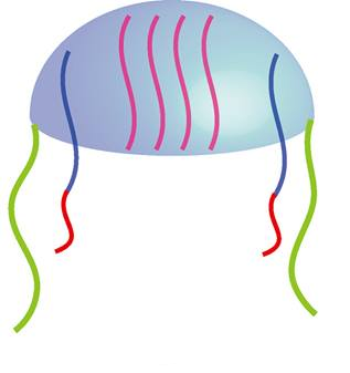
A
The H5 aptamers on the rim of the head (light green) play the role of the lock. Remember that there are 3 kinds of H5 aptamers, here, we use one of them (ATGACTTCTCCTAGTCAGACGGGTAACGTATGTTTTACA
TTACGAAATTTAGAGCACCCTTACAGCGAGACTCGTTGACCT
GTAGCAGTG)
. The H5 aptamers in the concave (pink) are different from the lock (sequence). Finally, there are long strands in the middle of the head (blue and red). They are composed of 2 parts: poly-C on the inner side and the complementary strand of the inner H5 aptamers.
<img src="http://openwetware.org/images/f/fd/Design3.png"/>
B
This kind of jellyfish only differ a little from the first one. The lock (yellow) isn’t H5 aptamers anymore, but the complementary strands of H5 aptamers instead. All other components are the same as the A jellyfish.
<img src="http://openwetware.org/images/3/34/Design4.png"/>
C
C jellyfish is similar to A, only the poly-C chain is changed to poly-G chain (dark green). The lock and the oligonucleotides inside the head are all the same as A.
<img src="http://openwetware.org/images/a/aa/Design2.png"/>
D
As with D, the only difference from C is that its lock is replaced by the complementary strands of the H5 aptamers (yellow). All other components are the same as C.
We have introduced all the components of the jellyfish. We will now see how it works.
<img src="http://openwetware.org/images/4/46/Designpic.jpg"/>
1.The four types of jellyfish are synthesized individually. The complementary strands of H5 aptamers (blue) in the hemisphere will hybridize with the inner H5 aptamers (pink). Consequently, the poly-C and poly-G chains are packaged within the heads.
2.Mix jellyfish A with B, and C with D. Add CAP37 simultaneously. CAP37 is a chemoattractant for monocytes². CAP37 can be replaced by other drugs that have the desired effect and the suitable size.
3.Since A jellyfish has H5 aptamers on its rim, and B jellyfish has complementary strands of H5 aptamers, these 2 jellyfish would become a dimer when they meet each other. The aptamers and complementary strands will hybridize and combine the two hemispheres. In addition, because CAP37 is freely suspended in the medium, they will be incorporated into the hemisphere and the dimer. As a result, we will be able to obtain dimers containing CAP37. The same applies for jellyfish C and D.
4.Without the presence of H5N1 viruses, the dimers remain locked. Once H5N1 viruses are present, the H5 aptamers (locks) on A and C jellyfish will bind to the viruses (instead of the complementary strands), thus opening the locks. The dimer dissociates into two monomers.
5.CAP37 is released, and the outer and inner H5 aptamers will attach to the surface of viruses. The poly-C and poly-G chains no longer kept within the hemisphere, since the H5 aptamers (inner ones) are no longer bound to their complementary strands. These free poly-C and poly-G chains are then able to undergo hybridization, and thus initiating aggregation.
In summary, the dimers are able to dissociate upon binding viruses, and subsequently release the packaged drugs and undergo aggregation. The significance of aggregation can be illustrated using the example of CAP37. Recall that CAP37 is a monocyte chemoattractant. Since CAP37 are released from the jellyfish dimer, the location with the highest concentration of CAP37 would coincide with where the virus concentration would also be the highest. Circulating monocytes would be attracted by released CAP37 to this site (with the jellyfish and the virus) and neutralize the virus. The efficiency in which the immune system clears virus infection would therefore be enhanced.
1.Wang, R., Zhao, J., Jiang, T., Kwon, Y., Lu, H., Jiao, P., ... Li, Y. (2013). Selection and characterization of DNA aptamers for use in detection of avian influenza virus H5N1. Journal of Virological Methods, 362-369. Retrieved September 4, 2014, from http://www.sciencedirect.com/science/article/pii/S0166093413000827
2.CAP37, a human neutrophil-derived chemotactic factor with monocyte specific activity.
Pereira HA1, Shafer WM, Pohl J, Martin LE, Spitznagel JK
The Journal of Clinical Investigation 1990 May;85(5):1468-76
</body>