IGEM:Harvard/2006/Adaptamers/Notebook/2006-7-18
7/18
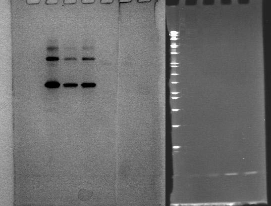
Ran a gel with multiple lanes of the same condition in order to have separate visualizations of DNA and protein.
Lanes are as follow:
2: BSA
3: thrombin in BSA
4: thrombin + T5
5: streptavidin
6: streptavidin + S5
7: streptavidin + biotinylated oligos
8: 1 kb ladder
9: 5S
10: 5S + strep
11: 5T
12: 5T + thromb
Where combinations are noted, 4 picomoles DNA + 2 picomoles protein were incubated for 30 minutes in a total reaction volume of 4 uL. Following incubations, solutions were diluted to 10uL including loading dye and run on a 12% polyacrylamide gel at 120 V for 1.5 hours.
Lanes 1-7 were stained with Coomassie Blue; lanes 8-12 with EtBr.
The lane with BSA clearly showed three bands, disagreeing with the results of the Nanostructure group, which suggested that only the bottom-most band belonged to BSA, and the others to thrombin. It was hoped that we had simply added thrombin to the BSA lane, but results from 7/21 showed that BSA in fact was responsible for all the bands.
No shift was observed for thrombin, although both DNA and aptamer bands appeared darker in lanes where they were incubated together, which is inconsistent with binding. Streptavidin only showed very lightly and not at all in the lane with biotin; perhaps streptavidin did bind biotin.
One curiosity is the bands stained with EtBr. S5 is more than double the length of T5, yet they appeared to migrate to the same location. I initially thought that the band somehow matched with the loading dye, but visual inspection showed that this was not the case.
