Organ-on-a-chip - Dan Nguyen
Background and Overview of an Organ-on-a-chip
Human organs are very complex units as each organ has different structures and contains different tissues and different types of cells. Traditional in vitro cell culture cannot yield satisfying results as it fails to correctly reconstitute a fully functional nature of a human body. Animal testings are often considered not accurate as well due to the animal models mostly fail to correctly predict the effects of drugs on humans. Organ-on-a-chip attempts to better fabricate the human organs, to hopefully, resolve the limitations that both 2-dimensional traditional cell culture methods and animal models bring about [1]. Organ-on-a-chip is a small-scaled multichannel 3-dimensional device that can imitate and reconstitute the microstructures and perform the functions of the living organs/tissues of a human body. To better imitate a complex human body environment, physiological conditions are taken into account while operating the devices such as fluid pH, cell culture medium, nutrient concentrations are monitored through pumps and valves in the organ-on-a-chip device. Different from the traditional cell culturing methods that are quiescent, organ-on-a-chips make use of fluid flows, as different fluid velocities, pressures, viscosities can affect on the nutrients/gases/cell concentration gradients, also act as mechanical stimuli to multiple cells present [2]. Creating an organ-on-a-chip is a very complicated task, as the human cellular environment is complex and ensuring cells proliferate and interact outside of its natural niches without contaminations is difficult.


Fabrication of an organ-on-a-chip
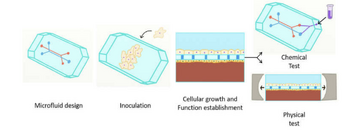
Different microfluidic devices that imitate lungs, kidneys, hearts or guts have been broadly researched. The general steps to create a specific organ on a chip are principally the same. The first steps are to design, mold and sterilize the device. Specific types of cells for the organ chips are perfused to the device with culture media through small inlets that are connected to small tubings. Cells are continuously grown inside the chip in a sterile environment. The model is often screened under the microscope for cell population check and once the cells divide enough, the chips will go through chemical tests such as cancer drug testing and drug screening [4].
Organ-on-a-chip applications
Possibilities for different organ-on-a-chips are creating drug delivery platforms for human specific-drug interactions
Integration of multiple organ-on-a-chip - Body-on-a-chip
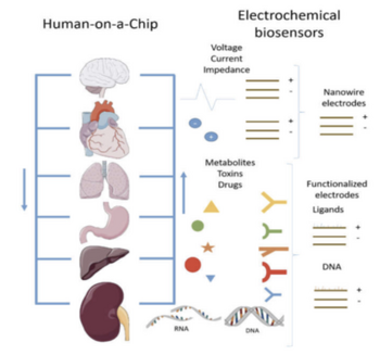
Once multiple organs-on-a-chips are extensively tested, and integration of multiple organ-on-a-chips would be the final goal to work on. Human-on-a-chip is one of the most important and most promising alternatives to animal testing as a human-on-a-chip is expected to perform as an entire human organ system (such as digestive system, respiratory system, cardiovascular system and so on) or even further, an entire living human. However, there are many challenges for the integration of all organ-on-a-chips because we have to ensure that once different chips are connected, they must support others like how human tissues support each other. Supporting computational models are needed to design an entire integration of organ-on-a-chips. In addition, developing a human-on-a-chip requires much more donated human cells and human materials (such as human bones) which are very expensive to get for research. Despite the challenges, human-on-a-chip is still a brilliant idea to be considered as once those human-on-a-chips become true, medical researches such as cancer treatment or drug testing no longer have to depend on animal testing.

Organ-on-a-chip advantages
Organ-on-a-chip is said to be promising as it attempts to imitate human organs that the traditional 2-D cell culturing methods and animal models fail to accurately predict. Organ-on-a-chip opens to many possibilities of combining drug testings, cell interaction visualization, microsensing of cell behaviors, that the other two traditional methods cannot achieve. The 3-dimensional shape, microstructures, and flexibility of the chips can mimic the human organ environment better. Organ-on-a-chip can also be combined with other techniques such as confocal microscopy, fluidic system that can optimize microchannels shapes that can enhance the nutrients and oxygen delivery to the cells. Another advantage that microfluidic chips hold is that crucial factors which are parts of the cell nurturing environment can be controlled and changed for testing for different purposes. Simple designs of microfluidics chips for simple simulations are easily made, the materials to make the chips are inexpensive (except human bones and human cells but those can be replaced with mice’s femur bones and cells or other animals’ bones and cells). Further cancer chemotherapy can be tested with microfluidics chips as drugs can also be inserted into the niches along with nutrients inside the culturing medium [3].
Organ-on-a-chip disadvantages
Alongside with the promising advantages, microfluidic chips may not reconstitute entire human organs as there are many other biomolecules inside the human body that are not available to insert in the microfluidics chips. The sizes of the microfluidics chips might not be to scale with the human organs and sizing matters because cells might behave differently with different volumes of fluids. Plus, microfluidics alone cannot sufficiently imitate every human organ. For example, heart-on-a-chip or nerves-on-a-chip are required to have some sort of electrochemical environment for precise simulations.
References
- Sosa-Hernández, J. E., Villalba-Rodríguez, A. M., Romero-Castillo, K. D., Aguilar-Aguila-Isaías, M. A., García-Reyes, I. E., Hernández-Antonio, A., . . . Iqbal, H. M. (2018). Organs-on-a-Chip Module: A Review from the Development and Applications Perspective. Micromachines, 9(10), 536. doi:10.3390/mi9100536
- Aziz, A., Geng, C., Fu, M., Yu, X., Qin, K., & Liu, B. (2017). The Role of Microfluidics for Organ on Chip Simulations, 4(4), 39. doi:10.3390/bioengineering4020039
- Marturano-Kruik, A., Nava, M. M., Yeager, K., Chramiec, A., Hao, L., Robinson, S., . . . Vunjak-Novakovic, G. (2018). Human bone perivascular niche-on-a-chip for studying metastatic colonization, 115(6), 1256-1261. doi:10.1073/pnas.1714282115
- Huh, D. (2015). A Human Breathing Lung-on-a-Chip, 12(1), S42-S44. doi:10.1513/annalsats.201410-442mg
- Yang, Q., Xu, F. (2017). Perspective: Fabrication of integrated organ-on-a-chip via bioprinting. Biomicrofluidics, 11(3). doi:10.1063/1.4982945
