Uploads by A. Florian Sessler
From OpenWetWare
Jump to navigationJump to search
This special page shows all uploaded files.
| Date | Name | Thumbnail | Size | Description |
|---|---|---|---|---|
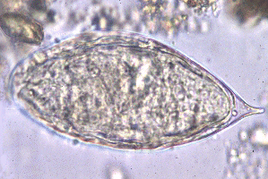
| 13:34, 21 October 2010 | Schistosoma egg.png (file) |  |
71 KB | Egg of S. ''haematobium''. [http://www.stanford.edu/class/humbio103/ParaSites2004/Schisto/website.html Stanford University (2004)] |
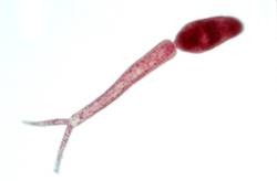
| 13:26, 21 October 2010 | Cercaria.jpg (file) |  |
4 KB | Cercaria [http://biology.unm.edu/biology/esloker/pi/SchistoEvolHistory.htm Loker Laboratory 2006] |
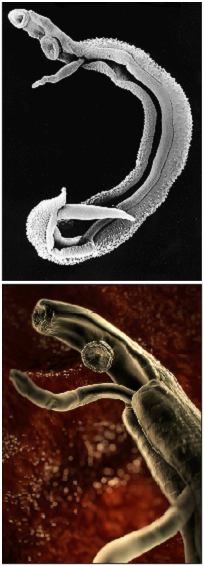
| 13:23, 21 October 2010 | Schistosoma2.png (file) |  |
145 KB | Pictures of schistosoma. Top: Electron microscopy of schistosoma couple [http://observationsofanerd.blogspot.com/2009/06/this-weeks-sci-fi-worthy-parasite.html Link]. Bottom: Artistic impression of schistosome parasite in blood vessel [http://animal.disco |
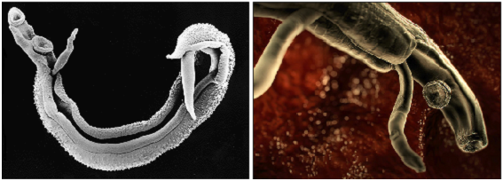
| 13:14, 21 October 2010 | Schistosoma.png (file) |  |
184 KB | Pictures of schistosoma. Left: Electron microscopy of schistosoma couple [http://observationsofanerd.blogspot.com/2009/06/this-weeks-sci-fi-worthy-parasite.html Link]. Right: Artistic impression of schistosome parasite in blood vessel [http://animal.disco |
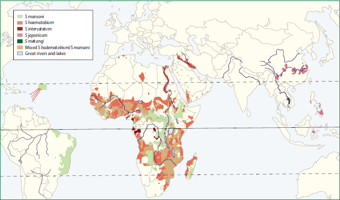
| 12:55, 21 October 2010 | Spread of schistosoma species (Gryseels et al. 2006).png (file) | .png) |
90 KB | Map of the distribution and spread of schistosoma species. (Gryseels et al. 2006) |
| 12:50, 21 October 2010 | Schistomes LifeCycle.gif (file) |  |
56 KB | Picture of the schistosoma life cycle. CDC (2010) |
| 16:09, 2 September 2010 | Harriett & florian head.JPG (file) | 24 KB | ||
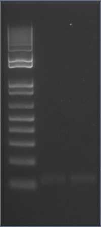
| 13:49, 2 September 2010 | 10-09-01 lytc psb1c3 purified for lig 2.jpg (file) |  |
6 KB | |
| 13:47, 2 September 2010 | 10-08-31 pSB1C3 pVEG1+2 concentration.jpg (file) |  |
9 KB | |
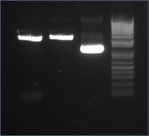
| 13:45, 2 September 2010 | 10-08-27 gel purification of pveg 50min.jpg (file) |  |
4 KB | |
| 13:43, 2 September 2010 | 10-08-25 diagnostic x+s s uncut psb12c3.jpg (file) |  |
9 KB | |
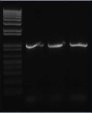
| 13:40, 2 September 2010 | 10-08-22 PCR CWB x 3.jpg (file) |  |
7 KB | |
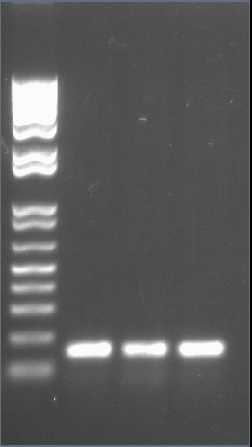
| 13:37, 2 September 2010 | 10-08-19 colony PCR 2.jpg (file) |  |
6 KB | |
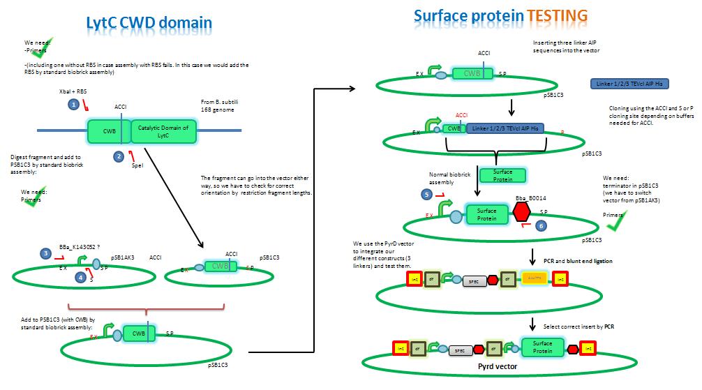
| 12:58, 2 September 2010 | Assembly LytC.JPG (file) |  |
70 KB | |
| 08:42, 31 August 2010 | Final background.JPG (file) |  |
38 KB | |
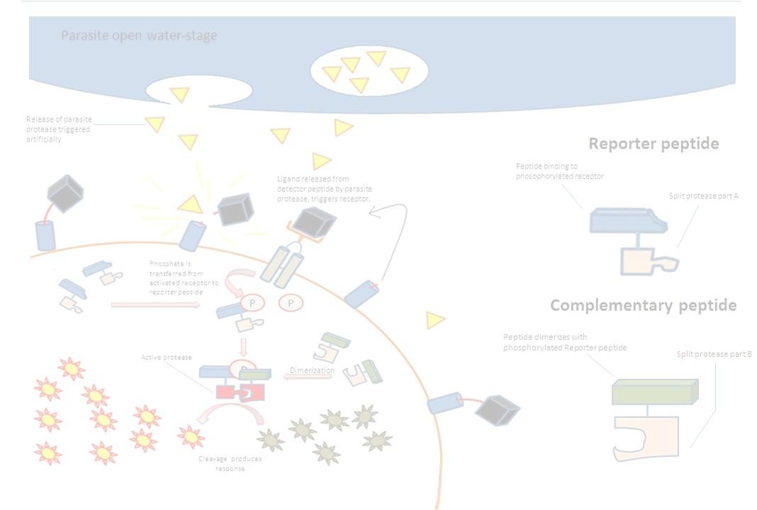
| 08:54, 29 August 2010 | Frm2.jpg (file) |  |
40 KB | background for the surface protein group |
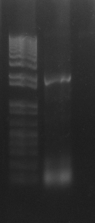
| 15:53, 16 August 2010 | 10-08-16 ligation reaction concs 4.bmp (file) |  |
138 KB | The last in a series of 4 pictures to estimate the concentration of our purified BOO14 and pSB1C3 |
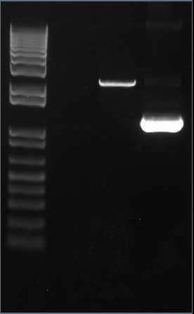
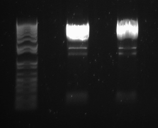
| 15:47, 16 August 2010 | 10-08-13 digests.bmp (file) |  |
243 KB | Gel confirming that our DNA (pSB1C3 and pSB1AK3 with BOO14) has been digested properly by PstI and EcoRI. |
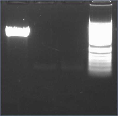
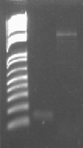
| 15:42, 16 August 2010 | 10-08-13 pSB1C3.bmp (file) |  |
240 KB | Gel of PCR confirming pSB1C3 has been amplified. |
| 15:12, 3 August 2010 | Table with membrane protein constructs.GIF (file) |  |
36 KB | Excel table with the fusion constructs we want to make to be put on the cell wall. |
| 15:23, 28 July 2010 | LytC sequences.docx (file) | 12 KB | ||
| 14:05, 21 July 2010 | Secretion system.GIF (file) |  |
22 KB | Protein secretion system as used by B. subtilis. |
| 13:59, 21 July 2010 | Table with secretion system molecules.GIF (file) |  |
19 KB | A table listing the components of the secretion system of B. subtilis. |
| 18:51, 15 July 2010 | Fatty acids.GIF (file) |  |
5 KB | |
| 14:34, 7 July 2010 | Fueller gross.jpg (file) |  |
18 KB | Füller |
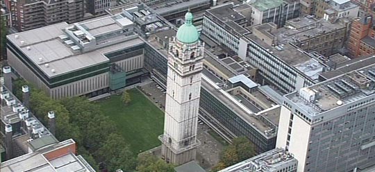
| 12:57, 7 July 2010 | Imperial campus.JPG (file) |  |
43 KB | Picture of Imperial College Campus |
