User:Senai H. Tesfamicael/Notebook/Biology 210 at AU
2/18/15 Zebrafish Experiment
Purpose: To understand the effects that the chemical nicotine has on the development of zebrafish embryos. The goal of understanding the effects of nicotine as reached by applying a testing group to an environment of nicotine applied through the water of their environment. The results and changes in development are then measured while being comparing them to the development of a control group of zebrafish.
Materials and Methods: The experiment required the use of 2 petri dishes one that would be used to hold the experimental group, containing the nicotine solution, and the other which would contain the control group of zebrafish. The petri dishes are then have twenty zebrafish added. The dishes are the periodically checked 3 times a eek to take measurements of the zebrafish development as ell as removing dead zebrafish, changing the water of the dishes, feeding the remaining fish.
Results: Treatment Date Hatched Alive Stage Movement Eye size (micrometers) Yolk size (micrometers) Notes 2/18/2015 0 20 18 hrs N/A 2/20/2015 6 20 36 slow movement possibly due to water 2/23/2015 10 14 72 hrs Shaking, doesn't move unless agitated 30 75 petruding mouth, full fin development 2/25/2015 0 10 (new) 60 hrs Fast, freely swims on it's own without agitation. Doesn't react to agitation 2.75 5.75 Started over, 10 new 2/27/2015 4 freely swims 3/2/2015 ALL DEAD; DIDNT START OVER
Control Date Hatched Alive Stage Movement Eye size (micrometers) Yolk size (micrometers) Notes 2/18/2015 0 20 18 hrs N/A 2/20/2015 8 18 36 slow movement possibly due to water 2/23/2015 5 6 48 hrs fast, freely moves, reacts to agitation 30 70 2/25/2015 1 6 72 hrs normal movement, responds to agitation 30 35 petruding mouth, full sized fins 2/27/2015 5 normal movment on most 30 30 3/2/2015 0 3 full grown shaking and spaztic 30 25
Conclusion: The conclusion that results from this experiment is that the nicotine had an fatal effect on the treatment group since no zebrafish had survived the experiment. This could also result from the experimental error on the part of the testers. The rate of development between the treatment group and the experimental group showed a disparity caused by the nicotine. This altogether shows that the were harmful to the development and life of the zebrafish.
2/10/15 Invertebrates Experiment
Purpose: To understand the characteristics of invertebrates in our individual transects of gain an understanding of the shape and specialized structures in the morphology of the invertebrates. We also aim to look at the movement patterns of the invertebrates.
Materials and Methods: Using a dissecting scope, the group observed the characteristics and methods of the acoelomates,psuedocoelomates, and coelomates. We attempted to look at the organs and muscular structures of the invertebrates.Secondly, example organisms were provided that allowed us to see examples of arachnida,diplopoda,chiplopoda, insect, and crustacea. Lastly, the Berlese funnel from last week experiment is used to gain invertebrates from our transects to observe.
Data: The movement of the acoelomates,psuedocoelomates, and coelomates were not able to be observed as the specimens are not alive. The structures of the cross sections of each are recorded however.
  Acoelomates(Planaria): Ectoderme,Mesoderme,Endoderme present-Simple 3-layered organism
  Psuedocoelomates (Nematode): 2 Ectoderme, 2 Mesoderme and a cavity section present- Multilayered organism
  Coelomates(Earthworm): Ectoderme, 2 Mesoderme, 2 Endoderme and a cavity section- Multilayered organism
Discussion: The organisms observed in dissecting scope varied in number of layers and size. The invertebrates observed from the Berlese funnel ranged in size and structure from anywhere of 10 mm to 1 cm. Typically, the larger organisms were the insects and the crustacean while the smaller organisms were the mites and nematodes.
1/21/15 Transect Hay Infusion Experiment
Purpose: To understand the characteristics, both abiotic and biotic, of my transect, the marsh, as ell as the gain an understanding of protists and algae' qualities. These ere done through observation of my transect alongside observation of protists microorganisms located in the transect soil.
Materials and Methods: Four Popsicle sticks are used to mark off a 20 20 transect to represent an environment. We then labeled the transect's abiotic and biotic factors while the group takes a drawing of the transect. Take a sample of the transect's soil in a new 50 ml container for our hay infusion culture. Then using the 10 ml of soil add 500 ml of water, 0.1 grams of dried milk, while gently stirring the mixture. Afterwards, leave the container open as to allow for oxygen.
Data: The transect, the marsh, contained manicured grass and area containing tall grasses and various plants which started dying as it is winter. The transect also contained concrete as the university constructed a sidewalk. The biotic factors of this transect also include the lawngrass, the various plants, and decomposed plant material left after dead plants. The abiotic factors of the transect include the soil, the rocks, boulders, concrete, snow , and various trash.
Conclusion The conclusion that I have obtained is the presence of protists in the transect and environment. I also discover the difference between the biotic factors and abiotic factors in the environment. Also,the role both of the biotic factors and abiotic factors on the environment. For example, the soil providing nutrients for the plants, abiotic, and the plants , Who decompose, provide the nutrients to the soil, biotic.
1/28/15 Bacteria Identification Lab
Purpose: To identify and comprehend the characteristics of bacteria found the in the assigned transect, the marsh. secondly, to identify and compare the various forms of bacteria characteristics like colony size, shape, method of motility, bacterium size, and presence of peptidoglycan membrane (gram positive or negative). This was done through observation of the bacteria, observation of bacterial colonies, and gram staining of the bacteria to determine presence of peptidoglycan.
Materials and Method: e began by inoculating our eight agar plates with samples from our hay infusion. Four of the agar contain just nutrients for the bacteria while the other four contain the nutrients and tetracycline, an antibiotic that certain bacteria could have resistance to. Using serial dilution, the amount of starting bacteria are a fraction of the previous to determine the growth rate. The bacteria are spread on the plates and left to breed. After growth, the bacteria are used for gram staining.
Data:
Dilution-10^-3 nutrient- 50 colonies-5.0*10^4 colonies/ml
Dilution-10^-5 nutrient- 61 colonies-6.1*10^6 colonies/ml
Dilution-10^-7 nutrient- 1 colony- 1*10^7 colonies/ml
Dilution-10^-9 nutrient- 1 colony- 1*10^9 colonies/ml
Dilution-10^-3 tetra.- 1 colony- 1*colony- 1*10^4 colonies/ml
Dilution-10^-5 tetra.- 56 colonies- 5.6*10^6 colonies/ml
Dilution-10^-7 tetra.- 0 colonies- 0 colonies/ml
Dilution-10^-9 tetra.- 0 colonies- 0 colonies/ml
Gram staining- All plates were positive for gram staining
Discussion: The transect contain gram positive bacteria, as well as various forms of bacteria except for archae bacteria which would not grow on these because of their specific requirements for habitat. The agar plates help to show that the tetracycline, on average, had an effect on the bacteria of our transect preventing growth or causing death of the organisms, with some growth by certain bacteria. The observation of certain bacteria showed various bacteria had different methods of movement while some had tails to propel them others had to extend their body to reach and pull. The agar plates showed that variation occurred hen looking at the size of colonies and color.
1/4/15 Plantae and Fungi Lab
Purpose: To understand the characteristics of the plants and fungi that are present in our individual transect, the marsh. e attempt to look at common characteristics like the presence of vascularization, method of reproduction, and presence of specialized structures.
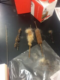
Materials and Method: Obtain 5 different plant samples from our transect. Take photos of the original samples. Take note of the height of the plant and the thickness of the stem. Take cross sections of the plants and look for the vascular tubing, xylem and phloem, and specialized structures like cuticle and stomata on leaves. Take notice of presence of seeds or other reproductive structures. Using a microscope, take an observation of fungi presence. Take a funnel filled with leaf litter from your transect over a flask of ethanol to catch invertebrates.
Data:
1. Seedless (Monocot)/ 3ft tall and thin/ Vascularized/ Asexual
2. Angiosperm (Monocot)/ Long thin with small flower located all around the stem/ Vascularized/ Asexual
3. Angiosperm (Monocot)/ 4 ft tall thick stem cotton like bush/ Vascularized/ Asexual
4. Angiosperm (Monocot) / short thin stem/ Vascularized/ Asexual
5. Angiosperm (Monocot)/ short thin stem/ Vascularized/ Asexual
 The plants are organized numerically with bottom being 1 and top being 5
Leaf litter large diversity w/ length ranging 1-3 inches and shapes ranging with some leaves with spreading outreaches and litter was in a compound pile 1 cm high.
Procedure VI: The fungus are stained in a blue color connected like nerve cells. It seems like it is part of the rhizopus and sprangia.
Discussion Observation of the transect showed that most of the collected plants were angiosperms with being of the seedless variety. The collected plants were all vascularized with tall stems approximately 2-4 ft in height. During this time of the year, there is a constant pile of leave litter located through a large portion of the marsh.